Manganese »
PDB 4ee3-4fo6 »
4ee3 »
Manganese in PDB 4ee3: Crystal Structure of Human M340H-Beta-1,4-Galactosyltransferase-1 (M340H-B4GAL-T1) in Complex with Pentasaccharide
Enzymatic activity of Crystal Structure of Human M340H-Beta-1,4-Galactosyltransferase-1 (M340H-B4GAL-T1) in Complex with Pentasaccharide
All present enzymatic activity of Crystal Structure of Human M340H-Beta-1,4-Galactosyltransferase-1 (M340H-B4GAL-T1) in Complex with Pentasaccharide:
2.4.1.22; 2.4.1.38; 2.4.1.90;
2.4.1.22; 2.4.1.38; 2.4.1.90;
Protein crystallography data
The structure of Crystal Structure of Human M340H-Beta-1,4-Galactosyltransferase-1 (M340H-B4GAL-T1) in Complex with Pentasaccharide, PDB code: 4ee3
was solved by
B.Ramakrishnan,
P.K.Qasba,
with X-Ray Crystallography technique. A brief refinement statistics is given in the table below:
| Resolution Low / High (Å) | 36.31 / 2.30 |
| Space group | C 2 2 21 |
| Cell size a, b, c (Å), α, β, γ (°) | 107.612, 195.158, 143.695, 90.00, 90.00, 90.00 |
| R / Rfree (%) | 18.7 / 24 |
Manganese Binding Sites:
The binding sites of Manganese atom in the Crystal Structure of Human M340H-Beta-1,4-Galactosyltransferase-1 (M340H-B4GAL-T1) in Complex with Pentasaccharide
(pdb code 4ee3). This binding sites where shown within
5.0 Angstroms radius around Manganese atom.
In total 3 binding sites of Manganese where determined in the Crystal Structure of Human M340H-Beta-1,4-Galactosyltransferase-1 (M340H-B4GAL-T1) in Complex with Pentasaccharide, PDB code: 4ee3:
Jump to Manganese binding site number: 1; 2; 3;
In total 3 binding sites of Manganese where determined in the Crystal Structure of Human M340H-Beta-1,4-Galactosyltransferase-1 (M340H-B4GAL-T1) in Complex with Pentasaccharide, PDB code: 4ee3:
Jump to Manganese binding site number: 1; 2; 3;
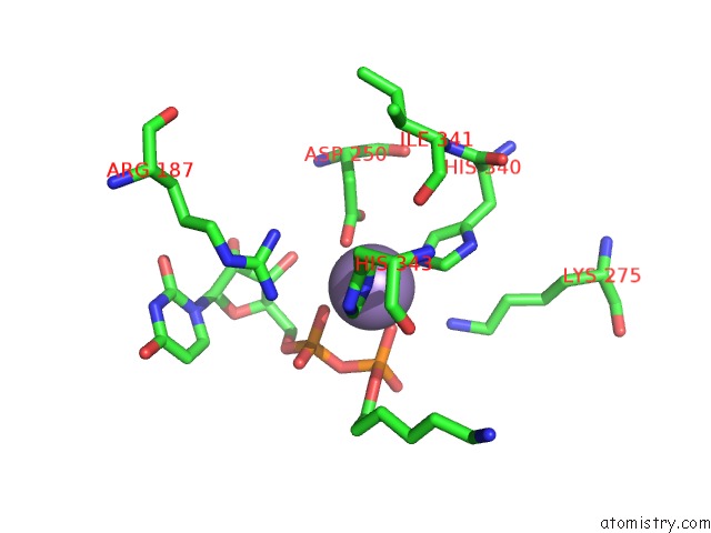
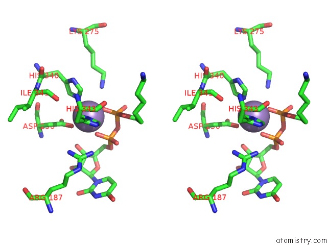
Manganese binding site 1 out of 3 in 4ee3
Go back to
Manganese binding site 1 out
of 3 in the Crystal Structure of Human M340H-Beta-1,4-Galactosyltransferase-1 (M340H-B4GAL-T1) in Complex with Pentasaccharide
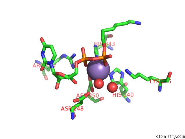
Mono view
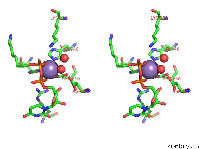
Stereo pair view
Mono view
Stereo pair view
A full contact list of Manganese with other atoms in the Mn binding
site number 1 of Crystal Structure of Human M340H-Beta-1,4-Galactosyltransferase-1 (M340H-B4GAL-T1) in Complex with Pentasaccharide within 5.0Å range:
|
Manganese binding site 2 out of 3 in 4ee3
Go back to
Manganese binding site 2 out
of 3 in the Crystal Structure of Human M340H-Beta-1,4-Galactosyltransferase-1 (M340H-B4GAL-T1) in Complex with Pentasaccharide
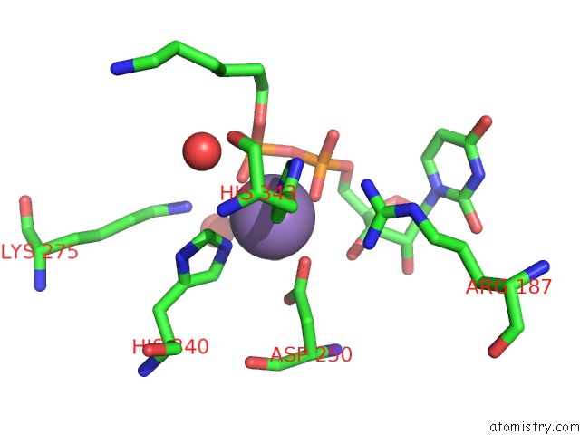
Mono view
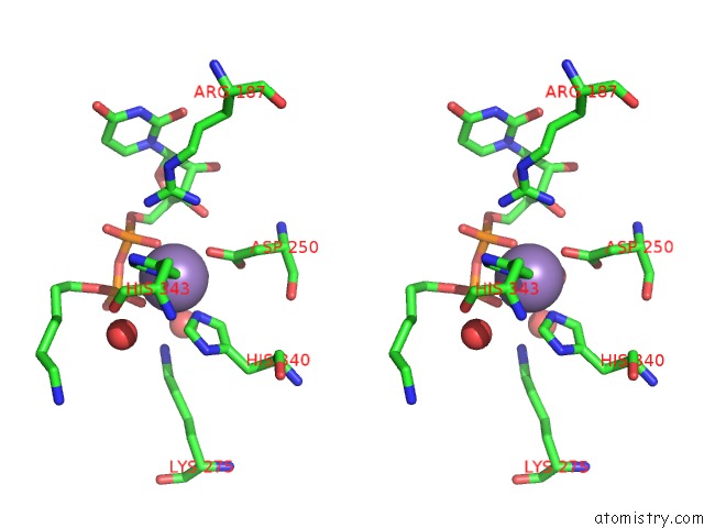
Stereo pair view
Mono view
Stereo pair view
A full contact list of Manganese with other atoms in the Mn binding
site number 2 of Crystal Structure of Human M340H-Beta-1,4-Galactosyltransferase-1 (M340H-B4GAL-T1) in Complex with Pentasaccharide within 5.0Å range:
|
Manganese binding site 3 out of 3 in 4ee3
Go back to
Manganese binding site 3 out
of 3 in the Crystal Structure of Human M340H-Beta-1,4-Galactosyltransferase-1 (M340H-B4GAL-T1) in Complex with Pentasaccharide
Mono view
Stereo pair view
Mono view
Stereo pair view
A full contact list of Manganese with other atoms in the Mn binding
site number 3 of Crystal Structure of Human M340H-Beta-1,4-Galactosyltransferase-1 (M340H-B4GAL-T1) in Complex with Pentasaccharide within 5.0Å range:
|
Reference:
B.Ramakrishnan,
E.Boeggeman,
P.K.Qasba.
Binding of N-Acetylglucosamine (Glcnac) Beta 1-6-Branched Oligosaccharide Acceptors to Beta 4-Galactosyltransferase I Reveals A New Ligand Binding Mode. J.Biol.Chem. V. 287 28666 2012.
ISSN: ISSN 0021-9258
PubMed: 22740701
DOI: 10.1074/JBC.M112.373514
Page generated: Sat Oct 5 19:18:57 2024
ISSN: ISSN 0021-9258
PubMed: 22740701
DOI: 10.1074/JBC.M112.373514
Last articles
Zn in 9J0NZn in 9J0O
Zn in 9J0P
Zn in 9FJX
Zn in 9EKB
Zn in 9C0F
Zn in 9CAH
Zn in 9CH0
Zn in 9CH3
Zn in 9CH1