Manganese »
PDB 4b5i-4dec »
4dec »
Manganese in PDB 4dec: Crystal Structure of Glucosyl-3-Phosphoglycerate Synthase From Mycobacterium Tuberculosis in Complex with MN2+, Uridine-Diphosphate (Udp) and Phosphoglyceric Acid (Pga)
Protein crystallography data
The structure of Crystal Structure of Glucosyl-3-Phosphoglycerate Synthase From Mycobacterium Tuberculosis in Complex with MN2+, Uridine-Diphosphate (Udp) and Phosphoglyceric Acid (Pga), PDB code: 4dec
was solved by
D.Albesa-Jove,
S.Urresti,
M.Van Der Woerd,
M.E.Guerin,
with X-Ray Crystallography technique. A brief refinement statistics is given in the table below:
| Resolution Low / High (Å) | 39.36 / 1.98 |
| Space group | I 41 |
| Cell size a, b, c (Å), α, β, γ (°) | 100.316, 100.316, 127.026, 90.00, 90.00, 90.00 |
| R / Rfree (%) | 17.7 / 20.5 |
Manganese Binding Sites:
The binding sites of Manganese atom in the Crystal Structure of Glucosyl-3-Phosphoglycerate Synthase From Mycobacterium Tuberculosis in Complex with MN2+, Uridine-Diphosphate (Udp) and Phosphoglyceric Acid (Pga)
(pdb code 4dec). This binding sites where shown within
5.0 Angstroms radius around Manganese atom.
In total only one binding site of Manganese was determined in the Crystal Structure of Glucosyl-3-Phosphoglycerate Synthase From Mycobacterium Tuberculosis in Complex with MN2+, Uridine-Diphosphate (Udp) and Phosphoglyceric Acid (Pga), PDB code: 4dec:
In total only one binding site of Manganese was determined in the Crystal Structure of Glucosyl-3-Phosphoglycerate Synthase From Mycobacterium Tuberculosis in Complex with MN2+, Uridine-Diphosphate (Udp) and Phosphoglyceric Acid (Pga), PDB code: 4dec:
Manganese binding site 1 out of 1 in 4dec
Go back to
Manganese binding site 1 out
of 1 in the Crystal Structure of Glucosyl-3-Phosphoglycerate Synthase From Mycobacterium Tuberculosis in Complex with MN2+, Uridine-Diphosphate (Udp) and Phosphoglyceric Acid (Pga)
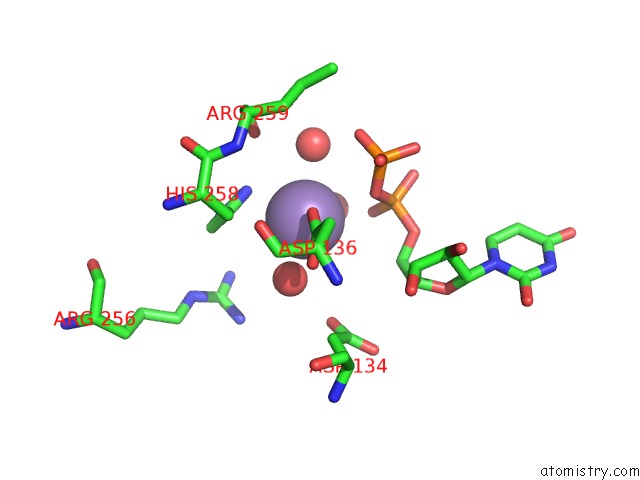
Mono view
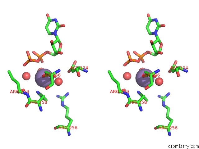
Stereo pair view
Mono view
Stereo pair view
A full contact list of Manganese with other atoms in the Mn binding
site number 1 of Crystal Structure of Glucosyl-3-Phosphoglycerate Synthase From Mycobacterium Tuberculosis in Complex with MN2+, Uridine-Diphosphate (Udp) and Phosphoglyceric Acid (Pga) within 5.0Å range:
|
Reference:
S.Urresti,
D.Albesa-Jove,
F.Schaeffer,
H.T.Pham,
D.Kaur,
P.Gest,
M.J.Van Der Woerd,
A.Carreras-Gonzalez,
S.Lopez-Fernandez,
P.M.Alzari,
P.J.Brennan,
M.Jackson,
M.E.Guerin.
Mechanistic Insights Into the Retaining Glucosyl-3-Phosphoglycerate Synthase From Mycobacteria. J.Biol.Chem. V. 287 24649 2012.
ISSN: ISSN 0021-9258
PubMed: 22637481
DOI: 10.1074/JBC.M112.368191
Page generated: Sat Oct 5 19:01:33 2024
ISSN: ISSN 0021-9258
PubMed: 22637481
DOI: 10.1074/JBC.M112.368191
Last articles
Zn in 9J0NZn in 9J0O
Zn in 9J0P
Zn in 9FJX
Zn in 9EKB
Zn in 9C0F
Zn in 9CAH
Zn in 9CH0
Zn in 9CH3
Zn in 9CH1