Manganese »
PDB 4b5i-4dec »
4ccl »
Manganese in PDB 4ccl: X-Ray Structure of E. Coli Ycfd
Protein crystallography data
The structure of X-Ray Structure of E. Coli Ycfd, PDB code: 4ccl
was solved by
M.A.Mcdonough,
C.H.Ho,
N.J.Kershaw,
C.J.Schofield,
with X-Ray Crystallography technique. A brief refinement statistics is given in the table below:
| Resolution Low / High (Å) | 41.765 / 2.60 |
| Space group | P 43 21 2 |
| Cell size a, b, c (Å), α, β, γ (°) | 120.693, 120.693, 133.498, 90.00, 90.00, 90.00 |
| R / Rfree (%) | 19.83 / 24.9 |
Manganese Binding Sites:
The binding sites of Manganese atom in the X-Ray Structure of E. Coli Ycfd
(pdb code 4ccl). This binding sites where shown within
5.0 Angstroms radius around Manganese atom.
In total 4 binding sites of Manganese where determined in the X-Ray Structure of E. Coli Ycfd, PDB code: 4ccl:
Jump to Manganese binding site number: 1; 2; 3; 4;
In total 4 binding sites of Manganese where determined in the X-Ray Structure of E. Coli Ycfd, PDB code: 4ccl:
Jump to Manganese binding site number: 1; 2; 3; 4;
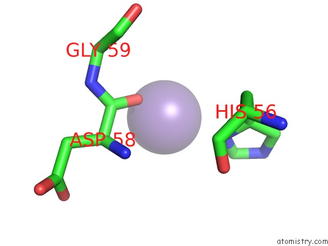
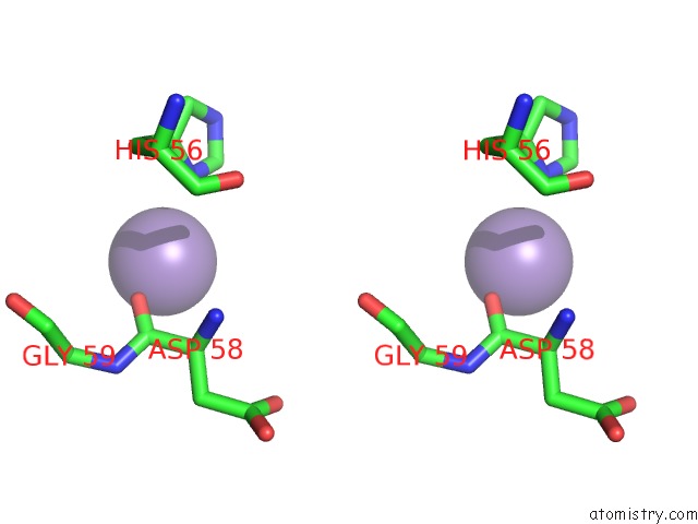
Manganese binding site 1 out of 4 in 4ccl
Go back to
Manganese binding site 1 out
of 4 in the X-Ray Structure of E. Coli Ycfd
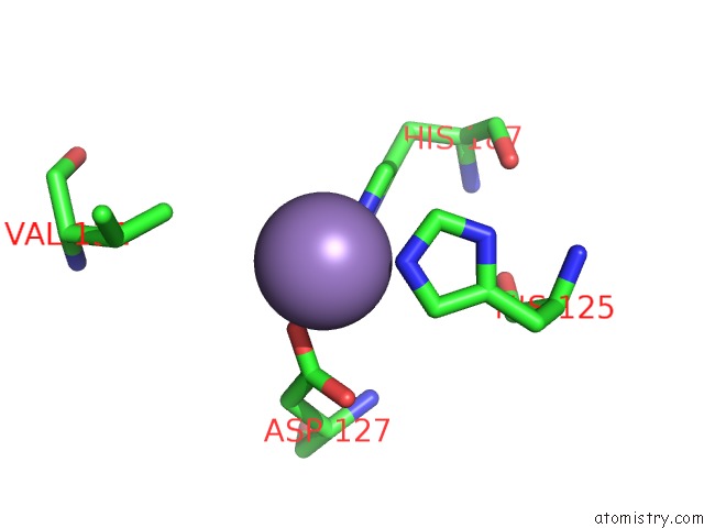
Mono view
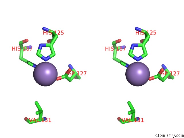
Stereo pair view
Mono view
Stereo pair view
A full contact list of Manganese with other atoms in the Mn binding
site number 1 of X-Ray Structure of E. Coli Ycfd within 5.0Å range:
|
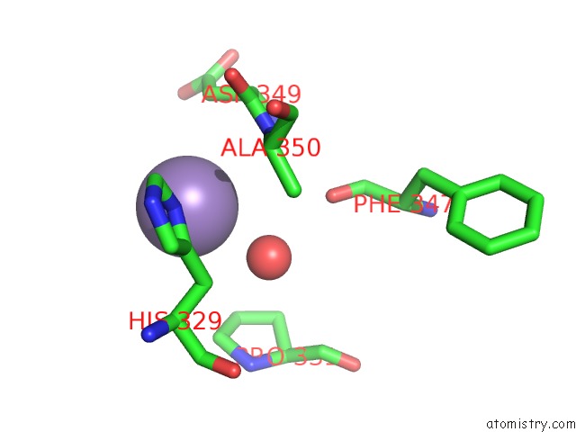
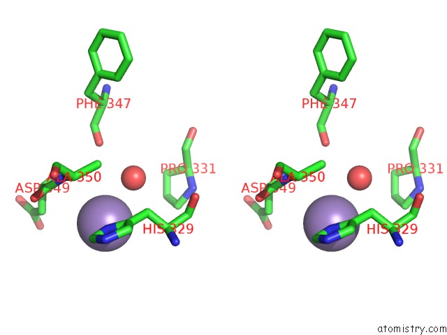
Manganese binding site 2 out of 4 in 4ccl
Go back to
Manganese binding site 2 out
of 4 in the X-Ray Structure of E. Coli Ycfd
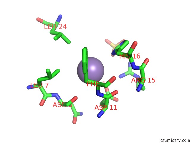
Mono view
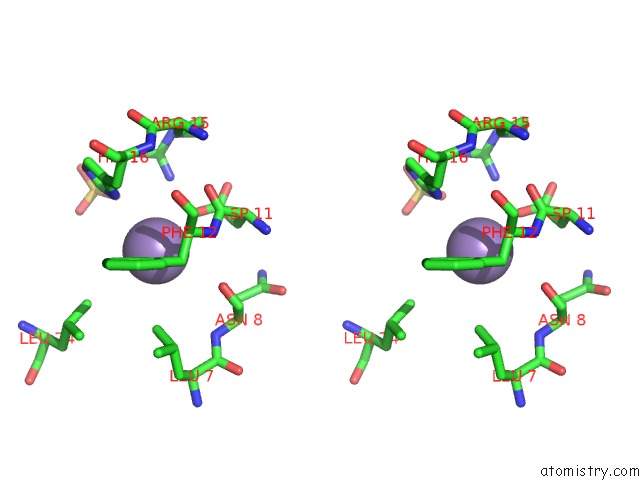
Stereo pair view
Mono view
Stereo pair view
A full contact list of Manganese with other atoms in the Mn binding
site number 2 of X-Ray Structure of E. Coli Ycfd within 5.0Å range:
|
Manganese binding site 3 out of 4 in 4ccl
Go back to
Manganese binding site 3 out
of 4 in the X-Ray Structure of E. Coli Ycfd
Mono view
Stereo pair view
Mono view
Stereo pair view
A full contact list of Manganese with other atoms in the Mn binding
site number 3 of X-Ray Structure of E. Coli Ycfd within 5.0Å range:
|
Manganese binding site 4 out of 4 in 4ccl
Go back to
Manganese binding site 4 out
of 4 in the X-Ray Structure of E. Coli Ycfd
Mono view
Stereo pair view
Mono view
Stereo pair view
A full contact list of Manganese with other atoms in the Mn binding
site number 4 of X-Ray Structure of E. Coli Ycfd within 5.0Å range:
|
Reference:
R.Chowdhury,
R.Sekirnik,
N.C.Brissett,
T.Krojer,
C.-H.Ho,
S.S.Ng,
I.J.Clifton,
W.Ge,
N.J.Kershaw,
G.C.Fox,
J.R.C.Muniz,
M.Vollmar,
C.Phillips,
E.S.Pilka,
K.L.Kavanagh,
F.Von Deflt,
U.Oppermann,
M.A.Mcdonough,
A.J.Doherty,
C.J.Schofield.
Ribosomal Oxygenases Are Structurally Conserved From Prokaryotes to Humans. Nature V. 510 422 2014.
ISSN: ISSN 0028-0836
PubMed: 24814345
DOI: 10.1038/NATURE13263
Page generated: Sat Oct 5 18:54:03 2024
ISSN: ISSN 0028-0836
PubMed: 24814345
DOI: 10.1038/NATURE13263
Last articles
F in 7K89F in 7K87
F in 7K6Z
F in 7K6M
F in 7K6A
F in 7K4D
F in 7K6L
F in 7K4F
F in 7K5M
F in 7K5E