Manganese »
PDB 3x30-4b5h »
4b5h »
Manganese in PDB 4b5h: Substate Bound Inactive Mutant of Neisseria Ap Endonuclease in Presence of Metal Ions
Enzymatic activity of Substate Bound Inactive Mutant of Neisseria Ap Endonuclease in Presence of Metal Ions
All present enzymatic activity of Substate Bound Inactive Mutant of Neisseria Ap Endonuclease in Presence of Metal Ions:
3.1.11.2;
3.1.11.2;
Protein crystallography data
The structure of Substate Bound Inactive Mutant of Neisseria Ap Endonuclease in Presence of Metal Ions, PDB code: 4b5h
was solved by
D.Lu,
J.Silhan,
J.T.Macdonald,
E.P.Carpenter,
K.Jensen,
C.M.Tang,
G.S.Baldwin,
P.S.Freemont,
with X-Ray Crystallography technique. A brief refinement statistics is given in the table below:
| Resolution Low / High (Å) | 31.07 / 3.05 |
| Space group | P 43 21 2 |
| Cell size a, b, c (Å), α, β, γ (°) | 68.907, 68.907, 120.968, 90.00, 90.00, 90.00 |
| R / Rfree (%) | 18.5 / 29.5 |
Manganese Binding Sites:
The binding sites of Manganese atom in the Substate Bound Inactive Mutant of Neisseria Ap Endonuclease in Presence of Metal Ions
(pdb code 4b5h). This binding sites where shown within
5.0 Angstroms radius around Manganese atom.
In total only one binding site of Manganese was determined in the Substate Bound Inactive Mutant of Neisseria Ap Endonuclease in Presence of Metal Ions, PDB code: 4b5h:
In total only one binding site of Manganese was determined in the Substate Bound Inactive Mutant of Neisseria Ap Endonuclease in Presence of Metal Ions, PDB code: 4b5h:
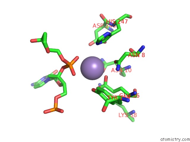
Manganese binding site 1 out of 1 in 4b5h
Go back to
Manganese binding site 1 out
of 1 in the Substate Bound Inactive Mutant of Neisseria Ap Endonuclease in Presence of Metal Ions
Mono view

Stereo pair view
Mono view
Stereo pair view
A full contact list of Manganese with other atoms in the Mn binding
site number 1 of Substate Bound Inactive Mutant of Neisseria Ap Endonuclease in Presence of Metal Ions within 5.0Å range:
|
Reference:
D.Lu,
J.Silhan,
J.T.Macdonald,
E.P.Carpenter,
K.Jensen,
C.M.Tang,
G.S.Baldwin,
P.S.Freemont.
Structural Basis For the Recognition and Cleavage of Abasic Dna in Neisseria Meningitidis. Proc. Natl. Acad. Sci. V. 109 16852 2012U.S.A..
ISSN: ESSN 1091-6490
PubMed: 23035246
DOI: 10.1073/PNAS.1206563109
Page generated: Sat Oct 5 18:48:51 2024
ISSN: ESSN 1091-6490
PubMed: 23035246
DOI: 10.1073/PNAS.1206563109
Last articles
Zn in 9MJ5Zn in 9HNW
Zn in 9G0L
Zn in 9FNE
Zn in 9DZN
Zn in 9E0I
Zn in 9D32
Zn in 9DAK
Zn in 8ZXC
Zn in 8ZUF