Manganese »
PDB 3vrs-3x2x »
3x1s »
Manganese in PDB 3x1s: Crystal Structure of the Nucleosome Core Particle
Protein crystallography data
The structure of Crystal Structure of the Nucleosome Core Particle, PDB code: 3x1s
was solved by
P.Sivaraman,
T.S.Kumarevel,
with X-Ray Crystallography technique. A brief refinement statistics is given in the table below:
| Resolution Low / High (Å) | 34.83 / 2.81 |
| Space group | P 21 21 21 |
| Cell size a, b, c (Å), α, β, γ (°) | 105.381, 109.363, 175.572, 90.00, 90.00, 90.00 |
| R / Rfree (%) | 22.1 / 27 |
Other elements in 3x1s:
The structure of Crystal Structure of the Nucleosome Core Particle also contains other interesting chemical elements:
| Chlorine | (Cl) | 2 atoms |
Manganese Binding Sites:
The binding sites of Manganese atom in the Crystal Structure of the Nucleosome Core Particle
(pdb code 3x1s). This binding sites where shown within
5.0 Angstroms radius around Manganese atom.
In total 6 binding sites of Manganese where determined in the Crystal Structure of the Nucleosome Core Particle, PDB code: 3x1s:
Jump to Manganese binding site number: 1; 2; 3; 4; 5; 6;
In total 6 binding sites of Manganese where determined in the Crystal Structure of the Nucleosome Core Particle, PDB code: 3x1s:
Jump to Manganese binding site number: 1; 2; 3; 4; 5; 6;
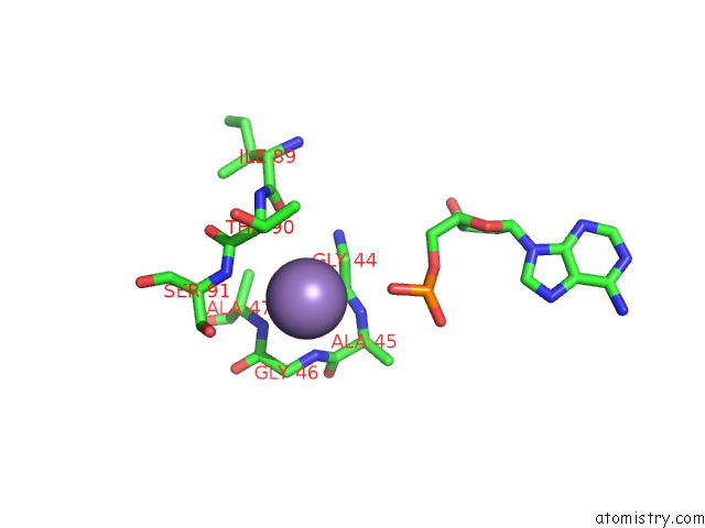
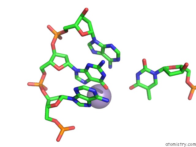
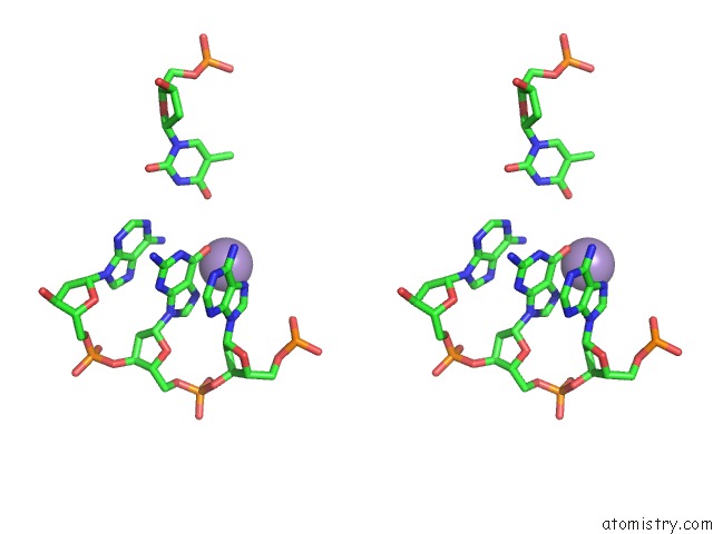
Manganese binding site 1 out of 6 in 3x1s
Go back to
Manganese binding site 1 out
of 6 in the Crystal Structure of the Nucleosome Core Particle
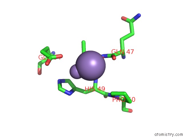
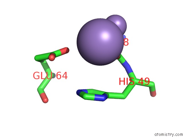
Mono view
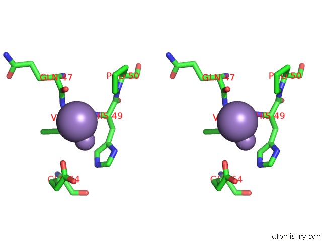
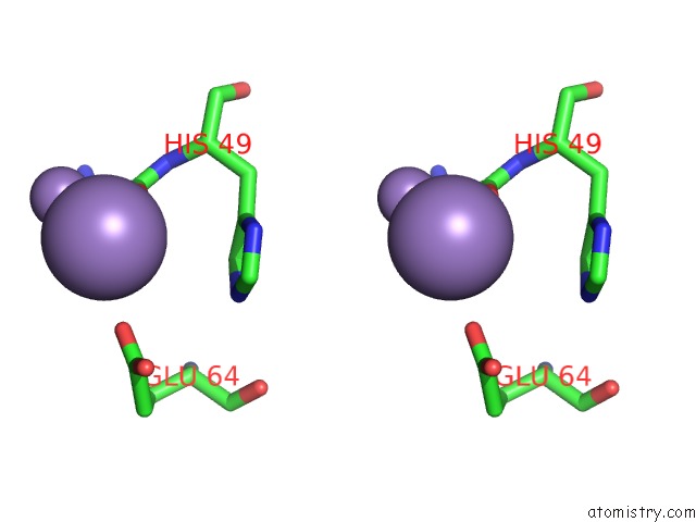
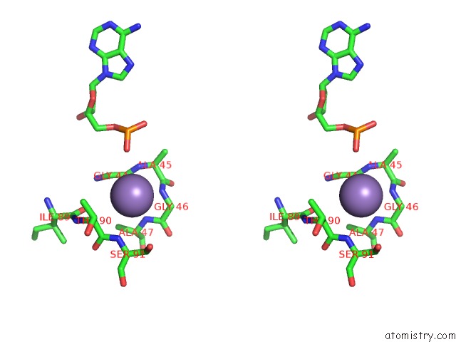
Stereo pair view
Mono view
Stereo pair view
A full contact list of Manganese with other atoms in the Mn binding
site number 1 of Crystal Structure of the Nucleosome Core Particle within 5.0Å range:
|
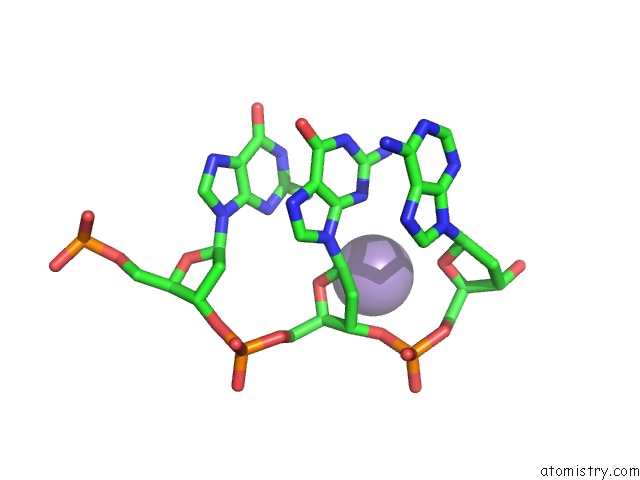
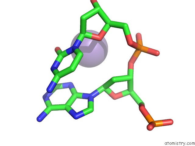
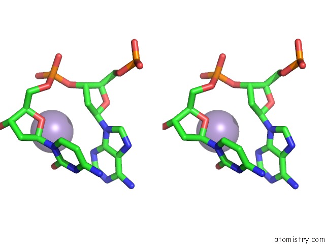
Manganese binding site 2 out of 6 in 3x1s
Go back to
Manganese binding site 2 out
of 6 in the Crystal Structure of the Nucleosome Core Particle
Mono view
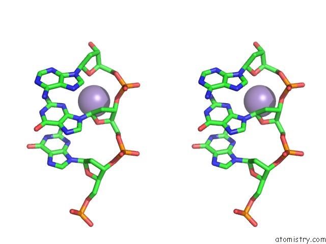
Stereo pair view
Mono view
Stereo pair view
A full contact list of Manganese with other atoms in the Mn binding
site number 2 of Crystal Structure of the Nucleosome Core Particle within 5.0Å range:
|
Manganese binding site 3 out of 6 in 3x1s
Go back to
Manganese binding site 3 out
of 6 in the Crystal Structure of the Nucleosome Core Particle
Mono view
Stereo pair view
Mono view
Stereo pair view
A full contact list of Manganese with other atoms in the Mn binding
site number 3 of Crystal Structure of the Nucleosome Core Particle within 5.0Å range:
|
Manganese binding site 4 out of 6 in 3x1s
Go back to
Manganese binding site 4 out
of 6 in the Crystal Structure of the Nucleosome Core Particle
Mono view
Stereo pair view
Mono view
Stereo pair view
A full contact list of Manganese with other atoms in the Mn binding
site number 4 of Crystal Structure of the Nucleosome Core Particle within 5.0Å range:
|
Manganese binding site 5 out of 6 in 3x1s
Go back to
Manganese binding site 5 out
of 6 in the Crystal Structure of the Nucleosome Core Particle
Mono view
Stereo pair view
Mono view
Stereo pair view
A full contact list of Manganese with other atoms in the Mn binding
site number 5 of Crystal Structure of the Nucleosome Core Particle within 5.0Å range:
|
Manganese binding site 6 out of 6 in 3x1s
Go back to
Manganese binding site 6 out
of 6 in the Crystal Structure of the Nucleosome Core Particle
Mono view
Stereo pair view
Mono view
Stereo pair view
A full contact list of Manganese with other atoms in the Mn binding
site number 6 of Crystal Structure of the Nucleosome Core Particle within 5.0Å range:
|
Reference:
S.Padavattan,
T.Shinagawa,
K.Hasegawa,
T.Kumasaka,
S.Ishii,
T.Kumarevel.
Structural and Functional Analyses of Nucleosome Complexes with Mouse Histone Variants TH2A and TH2B, Involved in Reprogramming Biochem.Biophys.Res.Commun. V. 464 929 2015.
ISSN: ISSN 0006-291X
PubMed: 26188507
DOI: 10.1016/J.BBRC.2015.07.070
Page generated: Sat Oct 5 18:33:33 2024
ISSN: ISSN 0006-291X
PubMed: 26188507
DOI: 10.1016/J.BBRC.2015.07.070
Last articles
Zn in 9J0NZn in 9J0O
Zn in 9J0P
Zn in 9FJX
Zn in 9EKB
Zn in 9C0F
Zn in 9CAH
Zn in 9CH0
Zn in 9CH3
Zn in 9CH1