Manganese »
PDB 3vrs-3x2x »
3wzh »
Manganese in PDB 3wzh: Crystal Structure of AFCSX3
Protein crystallography data
The structure of Crystal Structure of AFCSX3, PDB code: 3wzh
was solved by
Y.A.Yuan,
X.Yan,
with X-Ray Crystallography technique. A brief refinement statistics is given in the table below:
| Resolution Low / High (Å) | 22.88 / 3.31 |
| Space group | P 32 2 1 |
| Cell size a, b, c (Å), α, β, γ (°) | 86.726, 86.726, 112.529, 90.00, 90.00, 120.00 |
| R / Rfree (%) | 22.1 / 25.7 |
Manganese Binding Sites:
The binding sites of Manganese atom in the Crystal Structure of AFCSX3
(pdb code 3wzh). This binding sites where shown within
5.0 Angstroms radius around Manganese atom.
In total 5 binding sites of Manganese where determined in the Crystal Structure of AFCSX3, PDB code: 3wzh:
Jump to Manganese binding site number: 1; 2; 3; 4; 5;
In total 5 binding sites of Manganese where determined in the Crystal Structure of AFCSX3, PDB code: 3wzh:
Jump to Manganese binding site number: 1; 2; 3; 4; 5;
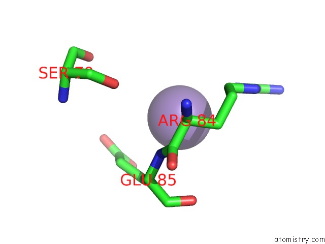
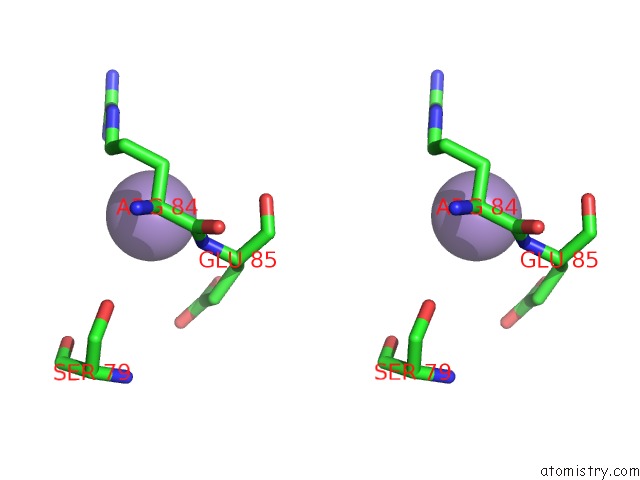
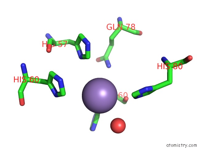
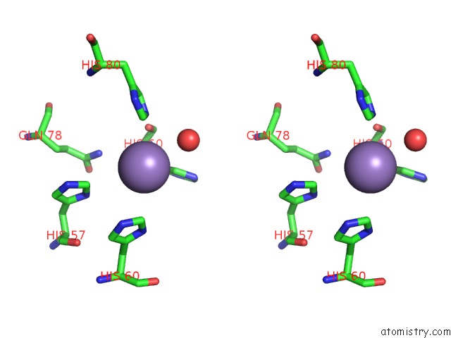
Manganese binding site 1 out of 5 in 3wzh
Go back to
Manganese binding site 1 out
of 5 in the Crystal Structure of AFCSX3
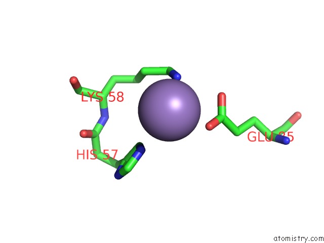
Mono view
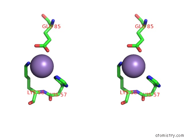
Stereo pair view
Mono view
Stereo pair view
A full contact list of Manganese with other atoms in the Mn binding
site number 1 of Crystal Structure of AFCSX3 within 5.0Å range:
|
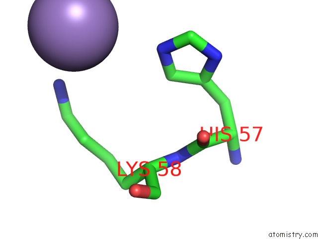
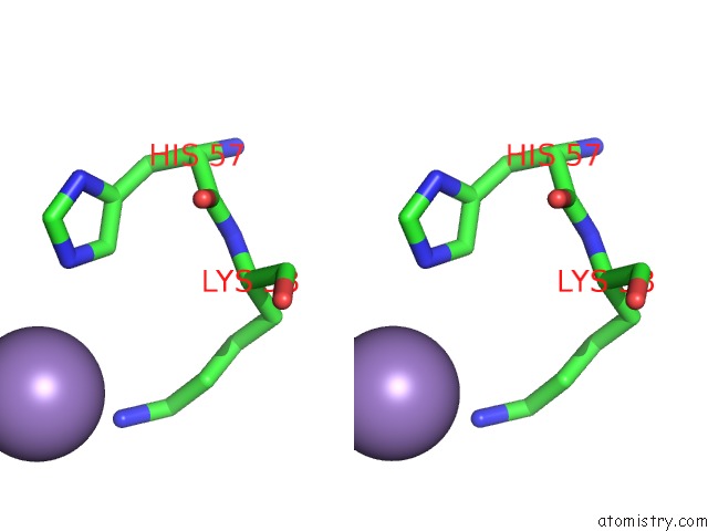
Manganese binding site 2 out of 5 in 3wzh
Go back to
Manganese binding site 2 out
of 5 in the Crystal Structure of AFCSX3
Mono view
Stereo pair view
Mono view
Stereo pair view
A full contact list of Manganese with other atoms in the Mn binding
site number 2 of Crystal Structure of AFCSX3 within 5.0Å range:
|
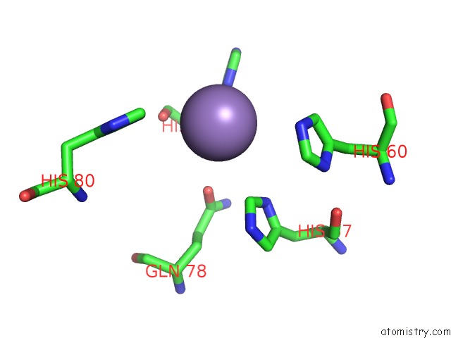
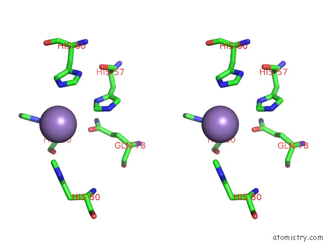
Manganese binding site 3 out of 5 in 3wzh
Go back to
Manganese binding site 3 out
of 5 in the Crystal Structure of AFCSX3
Mono view
Stereo pair view
Mono view
Stereo pair view
A full contact list of Manganese with other atoms in the Mn binding
site number 3 of Crystal Structure of AFCSX3 within 5.0Å range:
|
Manganese binding site 4 out of 5 in 3wzh
Go back to
Manganese binding site 4 out
of 5 in the Crystal Structure of AFCSX3
Mono view
Stereo pair view
Mono view
Stereo pair view
A full contact list of Manganese with other atoms in the Mn binding
site number 4 of Crystal Structure of AFCSX3 within 5.0Å range:
|
Manganese binding site 5 out of 5 in 3wzh
Go back to
Manganese binding site 5 out
of 5 in the Crystal Structure of AFCSX3
Mono view
Stereo pair view
Mono view
Stereo pair view
A full contact list of Manganese with other atoms in the Mn binding
site number 5 of Crystal Structure of AFCSX3 within 5.0Å range:
|
Reference:
X.Yan,
W.Guo,
Y.A.Yuan.
Crystal Structures of Crispr-Associated CSX3 Reveal A Manganese-Dependent Deadenylation Exoribonuclease Rna Biol. 2015.
ISSN: ESSN 1555-8584
PubMed: 26106927
DOI: 10.1080/15476286.2015.1051300
Page generated: Sat Oct 5 18:31:09 2024
ISSN: ESSN 1555-8584
PubMed: 26106927
DOI: 10.1080/15476286.2015.1051300
Last articles
Zn in 9J0NZn in 9J0O
Zn in 9J0P
Zn in 9FJX
Zn in 9EKB
Zn in 9C0F
Zn in 9CAH
Zn in 9CH0
Zn in 9CH3
Zn in 9CH1