Manganese »
PDB 3vrs-3x2x »
3ww1 »
Manganese in PDB 3ww1: X-Ray Structure of Cellulomonas Parahominis L-Ribose Isomerase with L- Ribose
Protein crystallography data
The structure of X-Ray Structure of Cellulomonas Parahominis L-Ribose Isomerase with L- Ribose, PDB code: 3ww1
was solved by
Y.Terami,
H.Yoshida,
G.Takata,
S.Kamitori,
with X-Ray Crystallography technique. A brief refinement statistics is given in the table below:
| Resolution Low / High (Å) | 19.33 / 1.95 |
| Space group | C 2 2 21 |
| Cell size a, b, c (Å), α, β, γ (°) | 76.730, 88.550, 152.020, 90.00, 90.00, 90.00 |
| R / Rfree (%) | 20.2 / 24.2 |
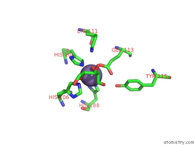
Manganese Binding Sites:
The binding sites of Manganese atom in the X-Ray Structure of Cellulomonas Parahominis L-Ribose Isomerase with L- Ribose
(pdb code 3ww1). This binding sites where shown within
5.0 Angstroms radius around Manganese atom.
In total 2 binding sites of Manganese where determined in the X-Ray Structure of Cellulomonas Parahominis L-Ribose Isomerase with L- Ribose, PDB code: 3ww1:
Jump to Manganese binding site number: 1; 2;
In total 2 binding sites of Manganese where determined in the X-Ray Structure of Cellulomonas Parahominis L-Ribose Isomerase with L- Ribose, PDB code: 3ww1:
Jump to Manganese binding site number: 1; 2;
Manganese binding site 1 out of 2 in 3ww1
Go back to
Manganese binding site 1 out
of 2 in the X-Ray Structure of Cellulomonas Parahominis L-Ribose Isomerase with L- Ribose

Mono view

Stereo pair view
Mono view
Stereo pair view
A full contact list of Manganese with other atoms in the Mn binding
site number 1 of X-Ray Structure of Cellulomonas Parahominis L-Ribose Isomerase with L- Ribose within 5.0Å range:
|
Manganese binding site 2 out of 2 in 3ww1
Go back to
Manganese binding site 2 out
of 2 in the X-Ray Structure of Cellulomonas Parahominis L-Ribose Isomerase with L- Ribose
Mono view

Stereo pair view
Mono view
Stereo pair view
A full contact list of Manganese with other atoms in the Mn binding
site number 2 of X-Ray Structure of Cellulomonas Parahominis L-Ribose Isomerase with L- Ribose within 5.0Å range:
|
Reference:
Y.Terami,
H.Yoshida,
K.Uechi,
K.Morimoto,
G.Takata,
S.Kamitori.
Essentiality of Tetramer Formation of Cellulomonas Parahominis L-Ribose Isomerase Involved in Novel L-Ribose Metabolic Pathway. Appl.Microbiol.Biotechnol. 2015.
ISSN: ESSN 1432-0614
PubMed: 25661811
DOI: 10.1007/S00253-015-6417-4
Page generated: Sat Oct 5 18:29:58 2024
ISSN: ESSN 1432-0614
PubMed: 25661811
DOI: 10.1007/S00253-015-6417-4
Last articles
Zn in 9MJ5Zn in 9HNW
Zn in 9G0L
Zn in 9FNE
Zn in 9DZN
Zn in 9E0I
Zn in 9D32
Zn in 9DAK
Zn in 8ZXC
Zn in 8ZUF