Manganese »
PDB 3vrs-3x2x »
3wcs »
Manganese in PDB 3wcs: Crystal Structure of Plant Lectin (Ligand-Bound Form)
Protein crystallography data
The structure of Crystal Structure of Plant Lectin (Ligand-Bound Form), PDB code: 3wcs
was solved by
M.Nagae,
Y.Yamaguchi,
with X-Ray Crystallography technique. A brief refinement statistics is given in the table below:
| Resolution Low / High (Å) | 98.10 / 1.75 |
| Space group | C 2 2 21 |
| Cell size a, b, c (Å), α, β, γ (°) | 76.579, 196.193, 98.655, 90.00, 90.00, 90.00 |
| R / Rfree (%) | 23.7 / 25.5 |
Other elements in 3wcs:
The structure of Crystal Structure of Plant Lectin (Ligand-Bound Form) also contains other interesting chemical elements:
| Calcium | (Ca) | 2 atoms |
Manganese Binding Sites:
The binding sites of Manganese atom in the Crystal Structure of Plant Lectin (Ligand-Bound Form)
(pdb code 3wcs). This binding sites where shown within
5.0 Angstroms radius around Manganese atom.
In total 2 binding sites of Manganese where determined in the Crystal Structure of Plant Lectin (Ligand-Bound Form), PDB code: 3wcs:
Jump to Manganese binding site number: 1; 2;
In total 2 binding sites of Manganese where determined in the Crystal Structure of Plant Lectin (Ligand-Bound Form), PDB code: 3wcs:
Jump to Manganese binding site number: 1; 2;
Manganese binding site 1 out of 2 in 3wcs
Go back to
Manganese binding site 1 out
of 2 in the Crystal Structure of Plant Lectin (Ligand-Bound Form)
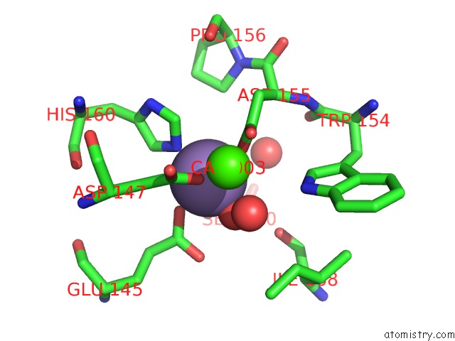
Mono view
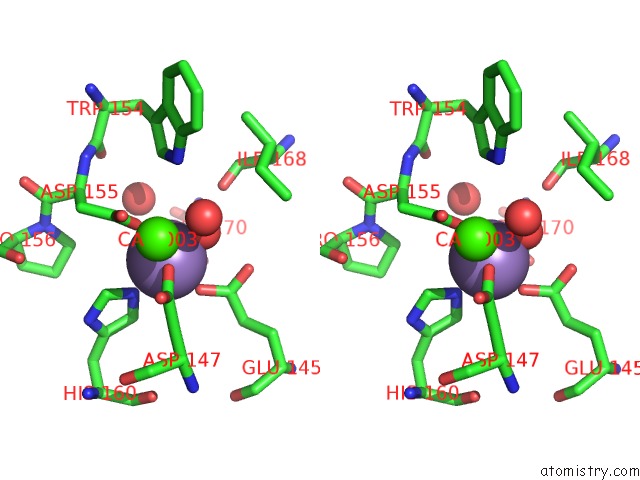
Stereo pair view
Mono view
Stereo pair view
A full contact list of Manganese with other atoms in the Mn binding
site number 1 of Crystal Structure of Plant Lectin (Ligand-Bound Form) within 5.0Å range:
|
Manganese binding site 2 out of 2 in 3wcs
Go back to
Manganese binding site 2 out
of 2 in the Crystal Structure of Plant Lectin (Ligand-Bound Form)
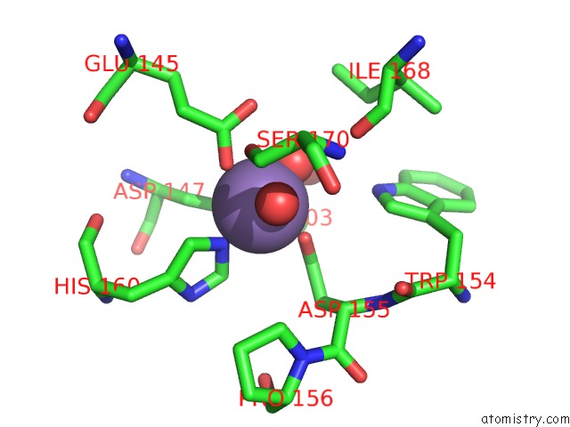
Mono view
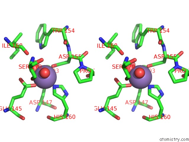
Stereo pair view
Mono view
Stereo pair view
A full contact list of Manganese with other atoms in the Mn binding
site number 2 of Crystal Structure of Plant Lectin (Ligand-Bound Form) within 5.0Å range:
|
Reference:
M.Nagae,
K.Soga,
K.Morita-Matsumoto,
S.Hanashima,
A.Ikeda,
K.Yamamoto,
Y.Yamaguchi.
Phytohemagglutinin From Phaseolus Vulgaris (Pha-E) Displays A Novel Glycan Recognition Mode Using A Common Legume Lectin Fold Glycobiology V. 24 368 2014.
ISSN: ISSN 0959-6658
PubMed: 24436051
DOI: 10.1093/GLYCOB/CWU004
Page generated: Sat Oct 5 18:26:24 2024
ISSN: ISSN 0959-6658
PubMed: 24436051
DOI: 10.1093/GLYCOB/CWU004
Last articles
Zn in 9J0NZn in 9J0O
Zn in 9J0P
Zn in 9FJX
Zn in 9EKB
Zn in 9C0F
Zn in 9CAH
Zn in 9CH0
Zn in 9CH3
Zn in 9CH1