Manganese »
PDB 3uag-3vnm »
3v1r »
Manganese in PDB 3v1r: Crystal Structures of the Reverse Transcriptase-Associated Ribonuclease H Domain of Xmrv with Inhibitor Beta-Thujaplicinol
Enzymatic activity of Crystal Structures of the Reverse Transcriptase-Associated Ribonuclease H Domain of Xmrv with Inhibitor Beta-Thujaplicinol
All present enzymatic activity of Crystal Structures of the Reverse Transcriptase-Associated Ribonuclease H Domain of Xmrv with Inhibitor Beta-Thujaplicinol:
3.1.26.4;
3.1.26.4;
Protein crystallography data
The structure of Crystal Structures of the Reverse Transcriptase-Associated Ribonuclease H Domain of Xmrv with Inhibitor Beta-Thujaplicinol, PDB code: 3v1r
was solved by
D.Zhou,
A.Wlodawer,
with X-Ray Crystallography technique. A brief refinement statistics is given in the table below:
| Resolution Low / High (Å) | 27.17 / 2.80 |
| Space group | C 2 2 21 |
| Cell size a, b, c (Å), α, β, γ (°) | 53.677, 84.750, 70.817, 90.00, 90.00, 90.00 |
| R / Rfree (%) | 19.5 / 27.7 |
Manganese Binding Sites:
The binding sites of Manganese atom in the Crystal Structures of the Reverse Transcriptase-Associated Ribonuclease H Domain of Xmrv with Inhibitor Beta-Thujaplicinol
(pdb code 3v1r). This binding sites where shown within
5.0 Angstroms radius around Manganese atom.
In total 2 binding sites of Manganese where determined in the Crystal Structures of the Reverse Transcriptase-Associated Ribonuclease H Domain of Xmrv with Inhibitor Beta-Thujaplicinol, PDB code: 3v1r:
Jump to Manganese binding site number: 1; 2;
In total 2 binding sites of Manganese where determined in the Crystal Structures of the Reverse Transcriptase-Associated Ribonuclease H Domain of Xmrv with Inhibitor Beta-Thujaplicinol, PDB code: 3v1r:
Jump to Manganese binding site number: 1; 2;
Manganese binding site 1 out of 2 in 3v1r
Go back to
Manganese binding site 1 out
of 2 in the Crystal Structures of the Reverse Transcriptase-Associated Ribonuclease H Domain of Xmrv with Inhibitor Beta-Thujaplicinol
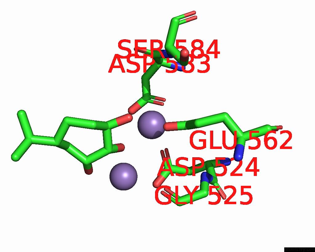
Mono view
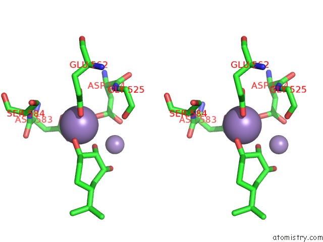
Stereo pair view
Mono view
Stereo pair view
A full contact list of Manganese with other atoms in the Mn binding
site number 1 of Crystal Structures of the Reverse Transcriptase-Associated Ribonuclease H Domain of Xmrv with Inhibitor Beta-Thujaplicinol within 5.0Å range:
|
Manganese binding site 2 out of 2 in 3v1r
Go back to
Manganese binding site 2 out
of 2 in the Crystal Structures of the Reverse Transcriptase-Associated Ribonuclease H Domain of Xmrv with Inhibitor Beta-Thujaplicinol
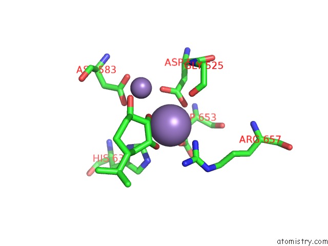
Mono view
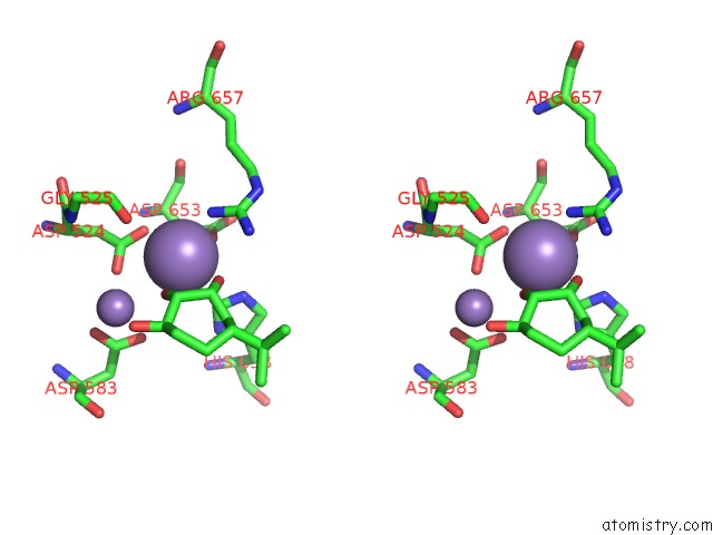
Stereo pair view
Mono view
Stereo pair view
A full contact list of Manganese with other atoms in the Mn binding
site number 2 of Crystal Structures of the Reverse Transcriptase-Associated Ribonuclease H Domain of Xmrv with Inhibitor Beta-Thujaplicinol within 5.0Å range:
|
Reference:
D.Zhou,
S.Chung,
M.Miller,
S.F.Le Grice,
A.Wlodawer.
Crystal Structures of the Reverse Transcriptase-Associated Ribonuclease H Domain of Xenotropic Murine Leukemia-Virus Related Virus. J.Struct.Biol. V. 177 638 2012.
ISSN: ISSN 1047-8477
PubMed: 22366278
DOI: 10.1016/J.JSB.2012.02.006
Page generated: Sat Oct 5 18:19:36 2024
ISSN: ISSN 1047-8477
PubMed: 22366278
DOI: 10.1016/J.JSB.2012.02.006
Last articles
Zn in 9J0NZn in 9J0O
Zn in 9J0P
Zn in 9FJX
Zn in 9EKB
Zn in 9C0F
Zn in 9CAH
Zn in 9CH0
Zn in 9CH3
Zn in 9CH1