Atomistry » Manganese » PDB 3uag-3vnm » 3uu0
Atomistry »
  Manganese »
    PDB 3uag-3vnm »
      3uu0 »

Manganese in PDB 3uu0: Crystal Structure of L-Rhamnose Isomerase From Bacillus Halodurans in Complex with Mn

Enzymatic activity of Crystal Structure of L-Rhamnose Isomerase From Bacillus Halodurans in Complex with Mn

All present enzymatic activity of Crystal Structure of L-Rhamnose Isomerase From Bacillus Halodurans in Complex with Mn:
5.3.1.14;

Protein crystallography data

The structure of Crystal Structure of L-Rhamnose Isomerase From Bacillus Halodurans in Complex with Mn, PDB code: 3uu0 was solved by T.T.N.Doan, P.Prabhu, J.K.Kim, M.Jeya, L.W.Kang, J.K.Lee, with X-Ray Crystallography technique. A brief refinement statistics is given in the table below:

Resolution Low / High (Å) 46.62 / 2.70
Space group P 1 21 1
Cell size a, b, c (Å), α, β, γ (°) 83.584, 165.553, 92.471, 90.00, 115.82, 90.00
R / Rfree (%) 18 / 25.5

Manganese Binding Sites:

The binding sites of Manganese atom in the Crystal Structure of L-Rhamnose Isomerase From Bacillus Halodurans in Complex with Mn (pdb code 3uu0). This binding sites where shown within 5.0 Angstroms radius around Manganese atom.
In total 8 binding sites of Manganese where determined in the Crystal Structure of L-Rhamnose Isomerase From Bacillus Halodurans in Complex with Mn, PDB code: 3uu0:
Jump to Manganese binding site number: 1; 2; 3; 4; 5; 6; 7; 8;

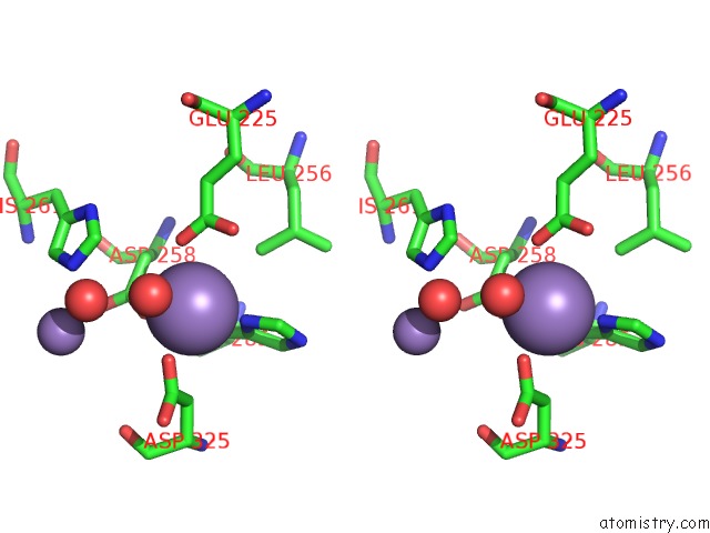
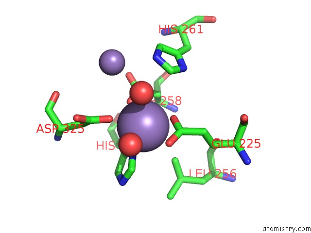
Manganese binding site 1 out of 8 in 3uu0

Go back to Manganese Binding Sites List in 3uu0
Manganese binding site 1 out of 8 in the Crystal Structure of L-Rhamnose Isomerase From Bacillus Halodurans in Complex with Mn


Mono view


Stereo pair view

A full contact list of Manganese with other atoms in the Mn binding site number 1 of Crystal Structure of L-Rhamnose Isomerase From Bacillus Halodurans in Complex with Mn within 5.0Å range:
probe atom residue distance (Å) B Occ
A:Mn419

b:45.8
occ:1.00
OD2 A:ASP258 2.0 29.0 1.0
OD2 A:ASP325 2.1 36.8 1.0
OE2 A:GLU225 2.2 27.7 1.0
ND1 A:HIS285 2.3 31.5 1.0
O A:HOH472 2.4 21.6 1.0
O A:HOH473 2.5 34.5 1.0
CG A:ASP258 3.1 25.5 1.0
CD A:GLU225 3.1 23.2 1.0
CE1 A:HIS285 3.2 32.0 1.0
CG A:ASP325 3.2 31.6 1.0
CG A:HIS285 3.3 29.1 1.0
OE1 A:GLU225 3.5 22.9 1.0
CB A:HIS285 3.6 28.1 1.0
CB A:ASP258 3.7 23.4 1.0
CB A:ASP325 3.8 30.2 1.0
CE1 A:HIS261 4.1 26.1 1.0
OD1 A:ASP258 4.2 27.4 1.0
OD1 A:ASP325 4.3 33.1 1.0
NE2 A:HIS285 4.3 31.5 1.0
CD2 A:HIS285 4.4 28.6 1.0
NE2 A:HIS261 4.4 26.6 1.0
CG A:GLU225 4.4 21.6 1.0
MN A:MN420 4.5 51.5 1.0
CD2 A:LEU256 4.8 21.9 1.0
ND1 A:HIS261 4.8 27.9 1.0
CA A:ASP258 4.8 23.6 1.0

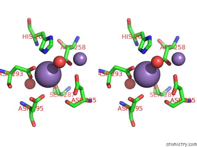
Manganese binding site 2 out of 8 in 3uu0

Go back to Manganese Binding Sites List in 3uu0
Manganese binding site 2 out of 8 in the Crystal Structure of L-Rhamnose Isomerase From Bacillus Halodurans in Complex with Mn


Mono view


Stereo pair view

A full contact list of Manganese with other atoms in the Mn binding site number 2 of Crystal Structure of L-Rhamnose Isomerase From Bacillus Halodurans in Complex with Mn within 5.0Å range:
probe atom residue distance (Å) B Occ
A:Mn420

b:51.5
occ:1.00
OD1 A:ASP293 1.9 28.6 1.0
OD2 A:ASP293 2.2 27.7 1.0
NE2 A:HIS261 2.3 26.6 1.0
CG A:ASP293 2.4 25.3 1.0
OD2 A:ASP295 2.9 36.0 1.0
CD2 A:HIS261 2.9 25.3 1.0
O A:HOH472 3.1 21.6 1.0
OD1 A:ASP258 3.4 27.4 1.0
CE1 A:HIS261 3.4 26.1 1.0
CG A:ASP295 3.7 32.1 1.0
OD1 A:ASP295 3.7 33.9 1.0
OD2 A:ASP258 3.7 29.0 1.0
CG A:ASP258 3.8 25.5 1.0
CB A:ASP293 3.9 22.5 1.0
O A:HOH469 4.1 35.5 1.0
CG A:HIS261 4.2 25.8 1.0
ND1 A:HIS261 4.3 27.9 1.0
OG A:SER287 4.4 25.7 1.0
MN A:MN419 4.5 45.8 1.0
NZ A:LYS227 4.6 27.8 1.0
OD2 A:ASP325 4.6 36.8 1.0
O A:ASP325 4.8 32.5 1.0
CA A:ASP293 4.8 23.7 1.0
CA A:GLY260 4.9 23.6 1.0
CB A:ASP258 4.9 23.4 1.0
CG A:ASP325 4.9 31.6 1.0
C A:GLY260 4.9 22.9 1.0
O A:GLY260 5.0 24.2 1.0

Manganese binding site 3 out of 8 in 3uu0

Go back to Manganese Binding Sites List in 3uu0
Manganese binding site 3 out of 8 in the Crystal Structure of L-Rhamnose Isomerase From Bacillus Halodurans in Complex with Mn


Mono view


Stereo pair view

A full contact list of Manganese with other atoms in the Mn binding site number 3 of Crystal Structure of L-Rhamnose Isomerase From Bacillus Halodurans in Complex with Mn within 5.0Å range:
probe atom residue distance (Å) B Occ
B:Mn419

b:37.3
occ:1.00
OD2 B:ASP325 2.3 35.4 1.0
ND1 B:HIS285 2.3 32.2 1.0
OD2 B:ASP258 2.3 24.7 1.0
OE2 B:GLU225 2.6 24.8 1.0
CG B:ASP325 3.2 29.1 1.0
CG B:HIS285 3.3 30.2 1.0
CE1 B:HIS285 3.3 33.1 1.0
CG B:ASP258 3.4 21.2 1.0
CD B:GLU225 3.4 24.4 1.0
CB B:ASP325 3.5 27.4 1.0
CB B:HIS285 3.5 27.0 1.0
OE1 B:GLU225 3.5 24.9 1.0
CB B:ASP258 3.8 20.6 1.0
O B:HOH471 4.1 37.8 1.0
CD1 B:LEU256 4.3 20.9 1.0
OD1 B:ASP325 4.4 27.2 1.0
NE2 B:HIS285 4.4 32.7 1.0
CE1 B:HIS261 4.4 21.5 1.0
CD2 B:HIS285 4.4 30.4 1.0
OD1 B:ASP258 4.5 20.4 1.0
O B:HOH477 4.6 43.7 1.0
NE2 B:HIS261 4.7 19.9 1.0
CG B:GLU225 4.7 24.3 1.0
MN B:MN420 4.9 63.7 1.0
CA B:ASP258 5.0 20.9 1.0
CA B:ASP325 5.0 28.0 1.0

Manganese binding site 4 out of 8 in 3uu0

Go back to Manganese Binding Sites List in 3uu0
Manganese binding site 4 out of 8 in the Crystal Structure of L-Rhamnose Isomerase From Bacillus Halodurans in Complex with Mn


Mono view


Stereo pair view

A full contact list of Manganese with other atoms in the Mn binding site number 4 of Crystal Structure of L-Rhamnose Isomerase From Bacillus Halodurans in Complex with Mn within 5.0Å range:
probe atom residue distance (Å) B Occ
B:Mn420

b:63.7
occ:1.00
OD2 B:ASP293 2.3 31.3 1.0
O B:HOH477 2.4 43.7 1.0
NE2 B:HIS261 2.6 19.9 1.0
OD1 B:ASP293 2.8 23.4 1.0
CG B:ASP293 2.9 25.3 1.0
OD1 B:ASP295 3.2 34.8 1.0
CD2 B:HIS261 3.4 19.0 1.0
CE1 B:HIS261 3.6 21.5 1.0
OD2 B:ASP258 3.9 24.7 1.0
CG B:ASP295 3.9 30.0 1.0
OD2 B:ASP295 4.0 35.0 1.0
OD2 B:ASP325 4.0 35.4 1.0
OD1 B:ASP258 4.3 20.4 1.0
CG B:ASP258 4.3 21.2 1.0
CB B:ASP293 4.3 22.5 1.0
O B:HOH471 4.5 37.8 1.0
CG B:ASP325 4.6 29.1 1.0
CG B:HIS261 4.6 20.0 1.0
NZ B:LYS227 4.7 28.3 1.0
O B:HOH442 4.7 17.4 1.0
ND1 B:HIS261 4.7 20.7 1.0
OG B:SER287 4.9 25.8 1.0
OD1 B:ASP325 4.9 27.2 1.0
MN B:MN419 4.9 37.3 1.0
O B:ASP293 5.0 22.1 1.0
O B:ASP325 5.0 31.6 1.0

Manganese binding site 5 out of 8 in 3uu0

Go back to Manganese Binding Sites List in 3uu0
Manganese binding site 5 out of 8 in the Crystal Structure of L-Rhamnose Isomerase From Bacillus Halodurans in Complex with Mn


Mono view


Stereo pair view

A full contact list of Manganese with other atoms in the Mn binding site number 5 of Crystal Structure of L-Rhamnose Isomerase From Bacillus Halodurans in Complex with Mn within 5.0Å range:
probe atom residue distance (Å) B Occ
C:Mn419

b:44.6
occ:1.00
OE1 C:GLU225 2.1 32.6 1.0
OD2 C:ASP325 2.3 35.9 1.0
OE2 C:GLU225 2.3 29.8 1.0
ND1 C:HIS285 2.4 28.8 1.0
O C:HOH476 2.5 26.8 1.0
CD C:GLU225 2.5 29.4 1.0
OD2 C:ASP258 2.6 30.9 1.0
CE1 C:HIS285 3.1 27.9 1.0
O C:HOH475 3.2 36.7 1.0
CG C:ASP325 3.5 31.4 1.0
CG C:HIS285 3.6 27.0 1.0
CG C:ASP258 3.6 22.9 1.0
CB C:ASP258 3.9 19.7 1.0
CG C:GLU225 4.0 25.7 1.0
CB C:HIS285 4.0 28.0 1.0
CE1 C:HIS261 4.1 28.8 1.0
CD2 C:LEU256 4.2 24.1 1.0
NE2 C:HIS285 4.3 26.3 1.0
OD1 C:ASP325 4.4 35.6 1.0
CB C:ASP325 4.4 27.9 1.0
CD2 C:HIS285 4.6 24.6 1.0
NE2 C:HIS261 4.6 28.9 1.0
MN C:MN420 4.7 53.3 1.0
OD1 C:ASP258 4.7 23.1 1.0
ND1 C:HIS261 4.8 28.6 1.0
CB C:GLU225 4.8 24.9 1.0

Manganese binding site 6 out of 8 in 3uu0

Go back to Manganese Binding Sites List in 3uu0
Manganese binding site 6 out of 8 in the Crystal Structure of L-Rhamnose Isomerase From Bacillus Halodurans in Complex with Mn


Mono view


Stereo pair view

A full contact list of Manganese with other atoms in the Mn binding site number 6 of Crystal Structure of L-Rhamnose Isomerase From Bacillus Halodurans in Complex with Mn within 5.0Å range:
probe atom residue distance (Å) B Occ
C:Mn420

b:53.3
occ:1.00
OD2 C:ASP293 2.0 31.1 1.0
CG C:ASP293 2.8 28.5 1.0
OD1 C:ASP293 2.8 28.3 1.0
NE2 C:HIS261 2.9 28.9 1.0
OD1 C:ASP295 3.0 37.4 1.0
OD1 C:ASP258 3.2 23.1 1.0
OD2 C:ASP258 3.3 30.9 1.0
CD2 C:HIS261 3.5 26.5 1.0
CG C:ASP258 3.5 22.9 1.0
OD2 C:ASP325 3.7 35.9 1.0
O C:HOH475 3.8 36.7 1.0
CG C:ASP295 3.8 32.4 1.0
OD2 C:ASP295 3.9 33.3 1.0
CE1 C:HIS261 3.9 28.8 1.0
CG C:ASP325 4.1 31.4 1.0
OG C:SER287 4.2 33.0 1.0
CB C:ASP293 4.3 26.3 1.0
OD1 C:ASP325 4.4 35.6 1.0
CG C:HIS261 4.6 25.7 1.0
MN C:MN419 4.7 44.6 1.0
O C:ASP325 4.7 29.2 1.0
O C:HOH434 4.8 32.3 1.0
CB C:ASP325 4.8 27.9 1.0
ND1 C:HIS261 4.8 28.6 1.0
CB C:ASP258 4.8 19.7 1.0
CB C:SER287 5.0 31.2 1.0

Manganese binding site 7 out of 8 in 3uu0

Go back to Manganese Binding Sites List in 3uu0
Manganese binding site 7 out of 8 in the Crystal Structure of L-Rhamnose Isomerase From Bacillus Halodurans in Complex with Mn


Mono view


Stereo pair view

A full contact list of Manganese with other atoms in the Mn binding site number 7 of Crystal Structure of L-Rhamnose Isomerase From Bacillus Halodurans in Complex with Mn within 5.0Å range:
probe atom residue distance (Å) B Occ
D:Mn419

b:50.8
occ:1.00
OD1 D:ASP258 2.0 27.2 1.0
O D:HOH478 2.3 30.0 1.0
ND1 D:HIS285 2.3 33.2 1.0
OE2 D:GLU225 2.4 32.2 1.0
OD1 D:ASP325 2.4 39.9 1.0
O D:HOH477 2.5 30.0 1.0
CD D:GLU225 3.2 29.0 1.0
CG D:ASP325 3.3 40.7 1.0
CG D:ASP258 3.3 26.1 1.0
CE1 D:HIS285 3.3 36.4 1.0
CG D:HIS285 3.3 31.6 1.0
OE1 D:GLU225 3.5 30.0 1.0
CB D:HIS285 3.6 30.5 1.0
CB D:ASP325 3.7 39.9 1.0
OD2 D:ASP258 4.1 29.0 1.0
CB D:ASP258 4.1 24.1 1.0
OD2 D:ASP325 4.2 45.5 1.0
CD1 D:LEU256 4.4 29.9 1.0
NE2 D:HIS285 4.4 36.6 1.0
CE1 D:HIS261 4.4 27.1 1.0
CD2 D:HIS285 4.5 34.1 1.0
CG D:GLU225 4.5 27.7 1.0
O D:HOH441 4.7 38.5 1.0
MN D:MN420 4.7 55.1 1.0
O D:HOH467 4.7 38.9 1.0
NE2 D:HIS261 4.8 28.3 1.0

Manganese binding site 8 out of 8 in 3uu0

Go back to Manganese Binding Sites List in 3uu0
Manganese binding site 8 out of 8 in the Crystal Structure of L-Rhamnose Isomerase From Bacillus Halodurans in Complex with Mn


Mono view


Stereo pair view

A full contact list of Manganese with other atoms in the Mn binding site number 8 of Crystal Structure of L-Rhamnose Isomerase From Bacillus Halodurans in Complex with Mn within 5.0Å range:
probe atom residue distance (Å) B Occ
D:Mn420

b:55.1
occ:1.00
OD2 D:ASP293 2.1 34.1 1.0
O D:HOH478 2.4 30.0 1.0
NE2 D:HIS261 2.4 28.3 1.0
OD1 D:ASP293 2.8 27.6 1.0
CG D:ASP293 2.8 29.1 1.0
O D:HOH467 2.9 38.9 1.0
OD1 D:ASP295 3.0 44.3 1.0
CD2 D:HIS261 3.4 26.1 1.0
CE1 D:HIS261 3.4 27.1 1.0
OD2 D:ASP295 3.5 41.7 1.0
CG D:ASP295 3.7 36.8 1.0
O D:HOH441 3.7 38.5 1.0
OD1 D:ASP325 3.9 39.9 1.0
OD1 D:ASP258 3.9 27.2 1.0
OD2 D:ASP258 4.1 29.0 1.0
CG D:ASP258 4.2 26.1 1.0
CB D:ASP293 4.3 27.2 1.0
NZ D:LYS227 4.4 28.3 1.0
ND1 D:HIS261 4.5 28.4 1.0
CG D:HIS261 4.5 25.9 1.0
O D:HOH477 4.6 30.0 1.0
MN D:MN419 4.7 50.8 1.0
CG D:ASP325 4.9 40.7 1.0
CD D:LYS227 4.9 27.0 1.0
OG D:SER287 5.0 30.0 1.0

Reference:

P.Prabhu, T.N.Doan, M.Tiwari, R.Singh, S.C.Kim, M.K.Hong, Y.C.Kang, L.W.Kang, J.K.Lee. Structure-Based Studies on the Metal Binding of Two-Metal-Dependent Sugar Isomerases. Febs J. V. 281 3446 2014.
ISSN: ISSN 1742-464X
PubMed: 24925069
DOI: 10.1111/FEBS.12872
Page generated: Tue Dec 15 04:16:29 2020

Last articles

Zn in 8WB0
Zn in 8WAX
Zn in 8WAU
Zn in 8WAZ
Zn in 8WAY
Zn in 8WAV
Zn in 8WAW
Zn in 8WAT
Zn in 8W7M
Zn in 8WD3
© Copyright 2008-2020 by atomistry.com
Home   |    Site Map   |    Copyright   |    Contact us   |    Privacy