Manganese »
PDB 3sx5-3u95 »
3u0y »
Manganese in PDB 3u0y: Crystal Structure of the Fucosylgalactoside Alpha N- Acetylgalactosaminyltransferase (Gta, Cisab Mutant L266G, G268A) in Complex with Compound 382 and Udp
Enzymatic activity of Crystal Structure of the Fucosylgalactoside Alpha N- Acetylgalactosaminyltransferase (Gta, Cisab Mutant L266G, G268A) in Complex with Compound 382 and Udp
All present enzymatic activity of Crystal Structure of the Fucosylgalactoside Alpha N- Acetylgalactosaminyltransferase (Gta, Cisab Mutant L266G, G268A) in Complex with Compound 382 and Udp:
2.4.1.37; 2.4.1.40;
2.4.1.37; 2.4.1.40;
Protein crystallography data
The structure of Crystal Structure of the Fucosylgalactoside Alpha N- Acetylgalactosaminyltransferase (Gta, Cisab Mutant L266G, G268A) in Complex with Compound 382 and Udp, PDB code: 3u0y
was solved by
M.M.Palcic,
R.Jorgensen,
with X-Ray Crystallography technique. A brief refinement statistics is given in the table below:
| Resolution Low / High (Å) | 19.86 / 1.60 |
| Space group | P 21 21 2 |
| Cell size a, b, c (Å), α, β, γ (°) | 77.630, 152.740, 52.990, 90.00, 90.00, 90.00 |
| R / Rfree (%) | 16.6 / 19.4 |
Manganese Binding Sites:
The binding sites of Manganese atom in the Crystal Structure of the Fucosylgalactoside Alpha N- Acetylgalactosaminyltransferase (Gta, Cisab Mutant L266G, G268A) in Complex with Compound 382 and Udp
(pdb code 3u0y). This binding sites where shown within
5.0 Angstroms radius around Manganese atom.
In total 2 binding sites of Manganese where determined in the Crystal Structure of the Fucosylgalactoside Alpha N- Acetylgalactosaminyltransferase (Gta, Cisab Mutant L266G, G268A) in Complex with Compound 382 and Udp, PDB code: 3u0y:
Jump to Manganese binding site number: 1; 2;
In total 2 binding sites of Manganese where determined in the Crystal Structure of the Fucosylgalactoside Alpha N- Acetylgalactosaminyltransferase (Gta, Cisab Mutant L266G, G268A) in Complex with Compound 382 and Udp, PDB code: 3u0y:
Jump to Manganese binding site number: 1; 2;
Manganese binding site 1 out of 2 in 3u0y
Go back to
Manganese binding site 1 out
of 2 in the Crystal Structure of the Fucosylgalactoside Alpha N- Acetylgalactosaminyltransferase (Gta, Cisab Mutant L266G, G268A) in Complex with Compound 382 and Udp
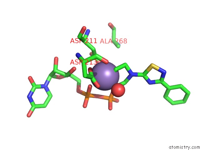
Mono view
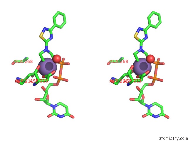
Stereo pair view
Mono view
Stereo pair view
A full contact list of Manganese with other atoms in the Mn binding
site number 1 of Crystal Structure of the Fucosylgalactoside Alpha N- Acetylgalactosaminyltransferase (Gta, Cisab Mutant L266G, G268A) in Complex with Compound 382 and Udp within 5.0Å range:
|
Manganese binding site 2 out of 2 in 3u0y
Go back to
Manganese binding site 2 out
of 2 in the Crystal Structure of the Fucosylgalactoside Alpha N- Acetylgalactosaminyltransferase (Gta, Cisab Mutant L266G, G268A) in Complex with Compound 382 and Udp
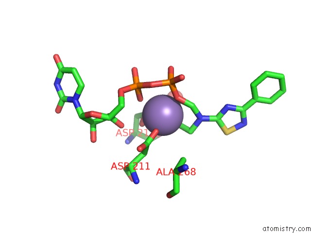
Mono view
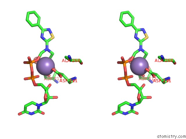
Stereo pair view
Mono view
Stereo pair view
A full contact list of Manganese with other atoms in the Mn binding
site number 2 of Crystal Structure of the Fucosylgalactoside Alpha N- Acetylgalactosaminyltransferase (Gta, Cisab Mutant L266G, G268A) in Complex with Compound 382 and Udp within 5.0Å range:
|
Reference:
R.Jorgensen,
L.L.Grimm,
N.Sindhuwinata,
T.Peters,
M.M.Palcic.
A Novel Compound From A Molecular Fragment Library Screen Inhibits Glycosyltransferases By Displacing the Metal Ion and Interfering with Substrate Binding To Be Published.
Page generated: Sat Oct 5 18:02:23 2024
Last articles
Zn in 9MJ5Zn in 9HNW
Zn in 9G0L
Zn in 9FNE
Zn in 9DZN
Zn in 9E0I
Zn in 9D32
Zn in 9DAK
Zn in 8ZXC
Zn in 8ZUF