Manganese »
PDB 3sx5-3u95 »
3taz »
Manganese in PDB 3taz: Crystal Structure of Nura Bound to Damp and Manganese
Protein crystallography data
The structure of Crystal Structure of Nura Bound to Damp and Manganese, PDB code: 3taz
was solved by
J.Chae,
Y.C.Kim,
Y.Cho,
with X-Ray Crystallography technique. A brief refinement statistics is given in the table below:
| Resolution Low / High (Å) | 28.71 / 3.20 |
| Space group | P 21 21 21 |
| Cell size a, b, c (Å), α, β, γ (°) | 65.158, 114.847, 123.414, 90.00, 90.00, 90.00 |
| R / Rfree (%) | 19.1 / 27.9 |
Manganese Binding Sites:
The binding sites of Manganese atom in the Crystal Structure of Nura Bound to Damp and Manganese
(pdb code 3taz). This binding sites where shown within
5.0 Angstroms radius around Manganese atom.
In total 2 binding sites of Manganese where determined in the Crystal Structure of Nura Bound to Damp and Manganese, PDB code: 3taz:
Jump to Manganese binding site number: 1; 2;
In total 2 binding sites of Manganese where determined in the Crystal Structure of Nura Bound to Damp and Manganese, PDB code: 3taz:
Jump to Manganese binding site number: 1; 2;
Manganese binding site 1 out of 2 in 3taz
Go back to
Manganese binding site 1 out
of 2 in the Crystal Structure of Nura Bound to Damp and Manganese
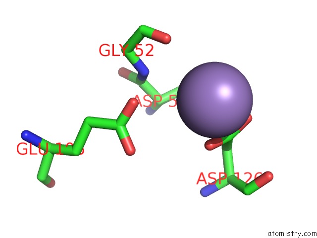
Mono view
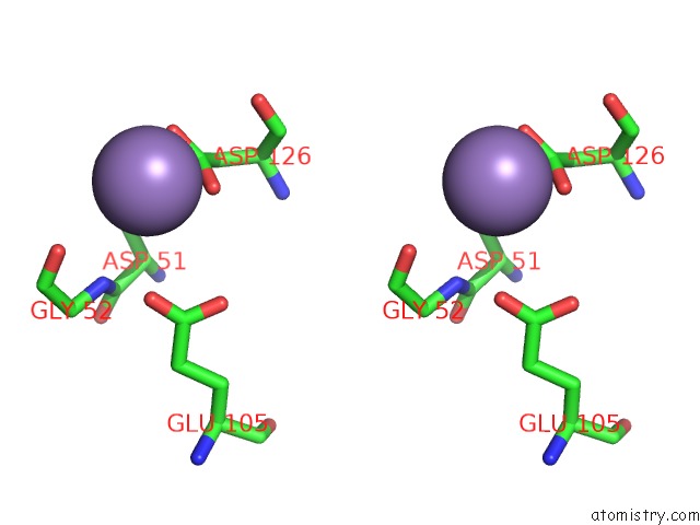
Stereo pair view
Mono view
Stereo pair view
A full contact list of Manganese with other atoms in the Mn binding
site number 1 of Crystal Structure of Nura Bound to Damp and Manganese within 5.0Å range:
|
Manganese binding site 2 out of 2 in 3taz
Go back to
Manganese binding site 2 out
of 2 in the Crystal Structure of Nura Bound to Damp and Manganese
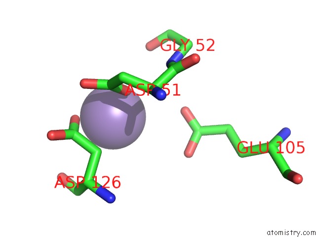
Mono view
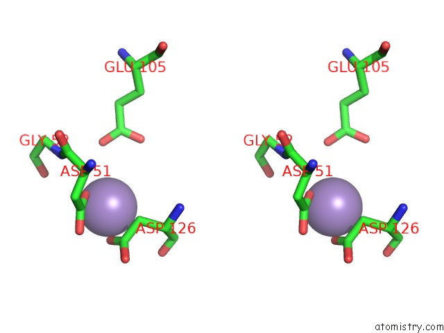
Stereo pair view
Mono view
Stereo pair view
A full contact list of Manganese with other atoms in the Mn binding
site number 2 of Crystal Structure of Nura Bound to Damp and Manganese within 5.0Å range:
|
Reference:
J.Chae,
Y.C.Kim,
Y.Cho.
Crystal Structure of the Nura-Damp-MN2+ Complex Nucleic Acids Res. V. 40 2258 2012.
ISSN: ISSN 0305-1048
PubMed: 22064858
DOI: 10.1093/NAR/GKR999
Page generated: Sat Oct 5 17:59:51 2024
ISSN: ISSN 0305-1048
PubMed: 22064858
DOI: 10.1093/NAR/GKR999
Last articles
Zn in 9J0NZn in 9J0O
Zn in 9J0P
Zn in 9FJX
Zn in 9EKB
Zn in 9C0F
Zn in 9CAH
Zn in 9CH0
Zn in 9CH3
Zn in 9CH1