Manganese »
PDB 3sx5-3u95 »
3tal »
Manganese in PDB 3tal: Crystal Structure of Nura with Manganese
Protein crystallography data
The structure of Crystal Structure of Nura with Manganese, PDB code: 3tal
was solved by
J.Chae,
Y.C.Kim,
Y.Cho,
with X-Ray Crystallography technique. A brief refinement statistics is given in the table below:
| Resolution Low / High (Å) | 29.48 / 3.15 |
| Space group | P 21 21 21 |
| Cell size a, b, c (Å), α, β, γ (°) | 64.803, 114.654, 121.624, 90.00, 90.00, 90.00 |
| R / Rfree (%) | 21.8 / 28.5 |
Manganese Binding Sites:
The binding sites of Manganese atom in the Crystal Structure of Nura with Manganese
(pdb code 3tal). This binding sites where shown within
5.0 Angstroms radius around Manganese atom.
In total 4 binding sites of Manganese where determined in the Crystal Structure of Nura with Manganese, PDB code: 3tal:
Jump to Manganese binding site number: 1; 2; 3; 4;
In total 4 binding sites of Manganese where determined in the Crystal Structure of Nura with Manganese, PDB code: 3tal:
Jump to Manganese binding site number: 1; 2; 3; 4;
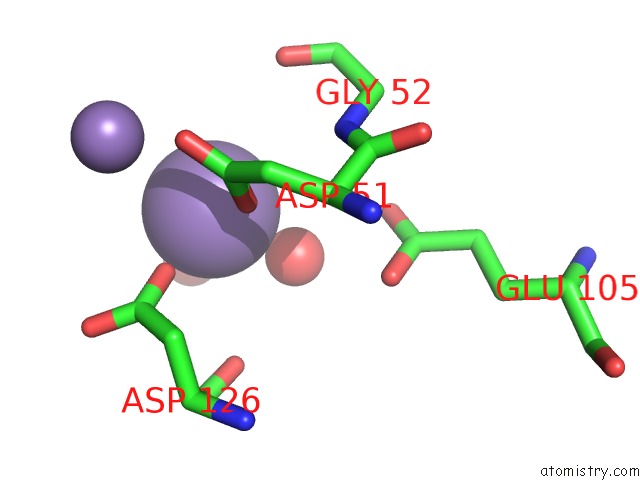
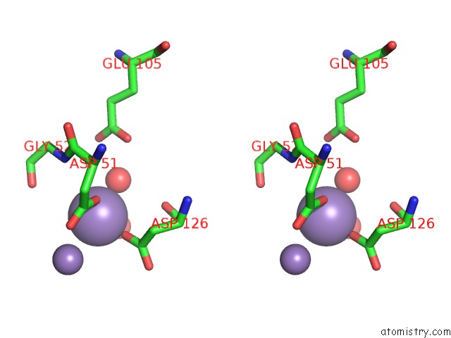
Manganese binding site 1 out of 4 in 3tal
Go back to
Manganese binding site 1 out
of 4 in the Crystal Structure of Nura with Manganese
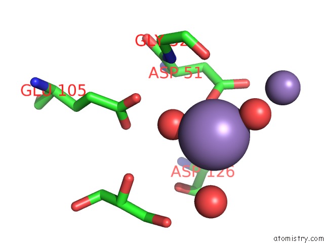
Mono view
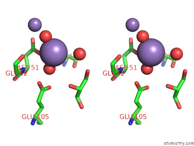
Stereo pair view
Mono view
Stereo pair view
A full contact list of Manganese with other atoms in the Mn binding
site number 1 of Crystal Structure of Nura with Manganese within 5.0Å range:
|
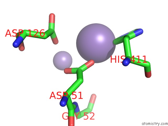
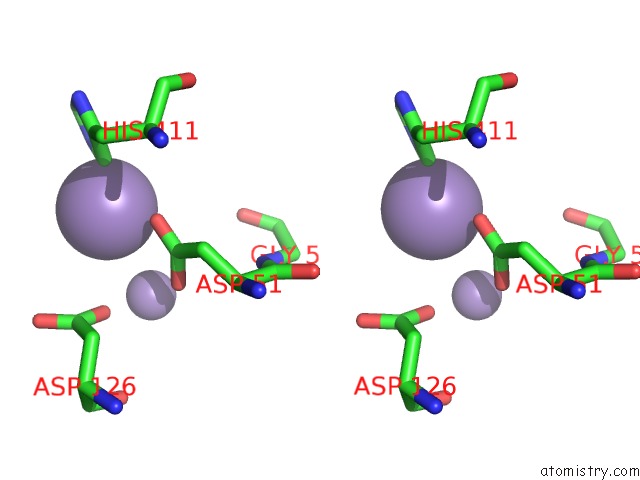
Manganese binding site 2 out of 4 in 3tal
Go back to
Manganese binding site 2 out
of 4 in the Crystal Structure of Nura with Manganese
Mono view
Stereo pair view
Mono view
Stereo pair view
A full contact list of Manganese with other atoms in the Mn binding
site number 2 of Crystal Structure of Nura with Manganese within 5.0Å range:
|
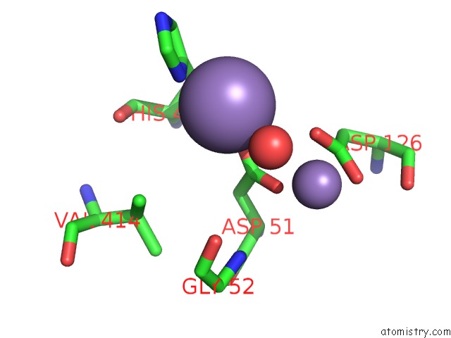
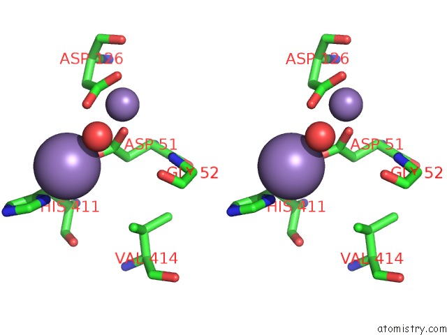
Manganese binding site 3 out of 4 in 3tal
Go back to
Manganese binding site 3 out
of 4 in the Crystal Structure of Nura with Manganese
Mono view
Stereo pair view
Mono view
Stereo pair view
A full contact list of Manganese with other atoms in the Mn binding
site number 3 of Crystal Structure of Nura with Manganese within 5.0Å range:
|
Manganese binding site 4 out of 4 in 3tal
Go back to
Manganese binding site 4 out
of 4 in the Crystal Structure of Nura with Manganese
Mono view
Stereo pair view
Mono view
Stereo pair view
A full contact list of Manganese with other atoms in the Mn binding
site number 4 of Crystal Structure of Nura with Manganese within 5.0Å range:
|
Reference:
J.Chae,
Y.C.Kim,
Y.Cho.
Crystal Structure of the Nura-Damp-MN2+ Complex Nucleic Acids Res. V. 40 2258 2012.
ISSN: ISSN 0305-1048
PubMed: 22064858
DOI: 10.1093/NAR/GKR999
Page generated: Sat Oct 5 17:59:31 2024
ISSN: ISSN 0305-1048
PubMed: 22064858
DOI: 10.1093/NAR/GKR999
Last articles
Cl in 3CZ0Cl in 3CYZ
Cl in 3CYW
Cl in 3CY2
Cl in 3CXW
Cl in 3CT9
Cl in 3CXP
Cl in 3CWL
Cl in 3CWC
Cl in 3CV7