Manganese »
PDB 3rla-3sx3 »
3s1s »
Manganese in PDB 3s1s: Characterization and Crystal Structure of the Type Iig Restriction Endonuclease Bpusi
Protein crystallography data
The structure of Characterization and Crystal Structure of the Type Iig Restriction Endonuclease Bpusi, PDB code: 3s1s
was solved by
B.W.Shen,
D.Xu,
S.-H.Chan,
Y.Zheng,
Y.Zhu,
S.-Y.Xu,
B.L.Stoddard,
with X-Ray Crystallography technique. A brief refinement statistics is given in the table below:
| Resolution Low / High (Å) | 30.00 / 2.35 |
| Space group | C 2 2 21 |
| Cell size a, b, c (Å), α, β, γ (°) | 213.472, 215.732, 73.710, 90.00, 90.00, 90.00 |
| R / Rfree (%) | 21.2 / 26.8 |
Other elements in 3s1s:
The structure of Characterization and Crystal Structure of the Type Iig Restriction Endonuclease Bpusi also contains other interesting chemical elements:
| Iodine | (I) | 38 atoms |
Manganese Binding Sites:
The binding sites of Manganese atom in the Characterization and Crystal Structure of the Type Iig Restriction Endonuclease Bpusi
(pdb code 3s1s). This binding sites where shown within
5.0 Angstroms radius around Manganese atom.
In total only one binding site of Manganese was determined in the Characterization and Crystal Structure of the Type Iig Restriction Endonuclease Bpusi, PDB code: 3s1s:
In total only one binding site of Manganese was determined in the Characterization and Crystal Structure of the Type Iig Restriction Endonuclease Bpusi, PDB code: 3s1s:
Manganese binding site 1 out of 1 in 3s1s
Go back to
Manganese binding site 1 out
of 1 in the Characterization and Crystal Structure of the Type Iig Restriction Endonuclease Bpusi
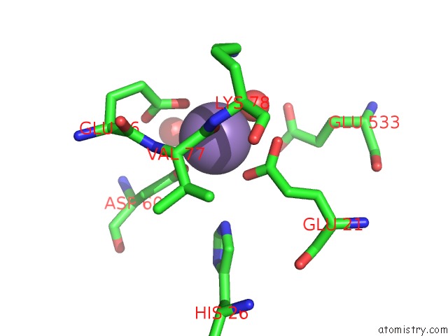
Mono view
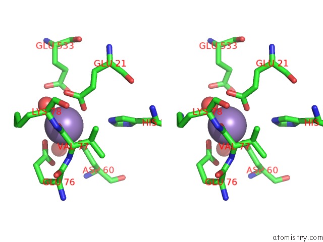
Stereo pair view
Mono view
Stereo pair view
A full contact list of Manganese with other atoms in the Mn binding
site number 1 of Characterization and Crystal Structure of the Type Iig Restriction Endonuclease Bpusi within 5.0Å range:
|
Reference:
B.W.Shen,
D.Xu,
S.H.Chan,
Y.Zheng,
Z.Zhu,
S.Y.Xu,
B.L.Stoddard.
Characterization and Crystal Structure of the Type Iig Restriction Endonuclease Rm.Bpusi. Nucleic Acids Res. V. 39 8223 2011.
ISSN: ISSN 0305-1048
PubMed: 21724614
DOI: 10.1093/NAR/GKR543
Page generated: Sat Oct 5 17:52:40 2024
ISSN: ISSN 0305-1048
PubMed: 21724614
DOI: 10.1093/NAR/GKR543
Last articles
Zn in 9J0NZn in 9J0O
Zn in 9J0P
Zn in 9FJX
Zn in 9EKB
Zn in 9C0F
Zn in 9CAH
Zn in 9CH0
Zn in 9CH3
Zn in 9CH1