Manganese »
PDB 3rla-3sx3 »
3rvo »
Manganese in PDB 3rvo: Structure of Chey-MN2+ Complex with Substitutions at 59 and 89: N59D E89Y
Protein crystallography data
The structure of Structure of Chey-MN2+ Complex with Substitutions at 59 and 89: N59D E89Y, PDB code: 3rvo
was solved by
R.M.Immormino,
C.A.Starbird,
R.E.Silversmith,
R.B.Bourret,
with X-Ray Crystallography technique. A brief refinement statistics is given in the table below:
| Resolution Low / High (Å) | 23.44 / 1.55 |
| Space group | P 21 21 21 |
| Cell size a, b, c (Å), α, β, γ (°) | 45.364, 46.885, 53.524, 90.00, 90.00, 90.00 |
| R / Rfree (%) | 15.2 / 18.5 |
Manganese Binding Sites:
The binding sites of Manganese atom in the Structure of Chey-MN2+ Complex with Substitutions at 59 and 89: N59D E89Y
(pdb code 3rvo). This binding sites where shown within
5.0 Angstroms radius around Manganese atom.
In total only one binding site of Manganese was determined in the Structure of Chey-MN2+ Complex with Substitutions at 59 and 89: N59D E89Y, PDB code: 3rvo:
In total only one binding site of Manganese was determined in the Structure of Chey-MN2+ Complex with Substitutions at 59 and 89: N59D E89Y, PDB code: 3rvo:
Manganese binding site 1 out of 1 in 3rvo
Go back to
Manganese binding site 1 out
of 1 in the Structure of Chey-MN2+ Complex with Substitutions at 59 and 89: N59D E89Y
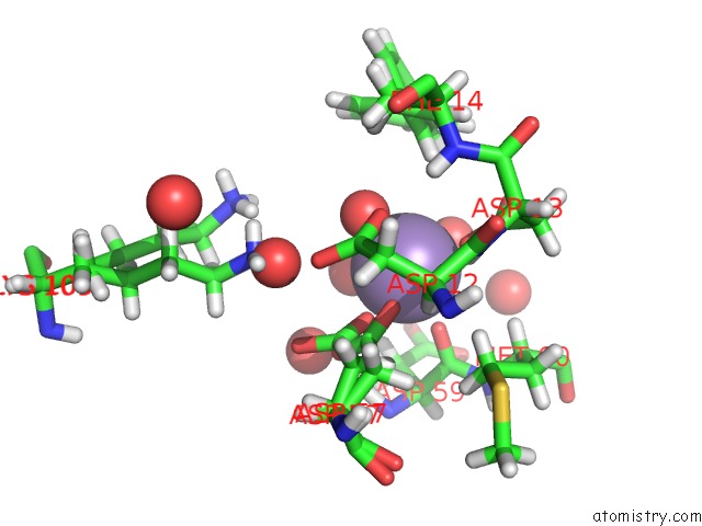
Mono view
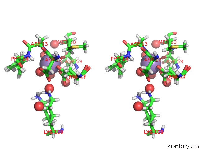
Stereo pair view
Mono view
Stereo pair view
A full contact list of Manganese with other atoms in the Mn binding
site number 1 of Structure of Chey-MN2+ Complex with Substitutions at 59 and 89: N59D E89Y within 5.0Å range:
|
Reference:
R.M.Immormino,
C.A.Starbird,
R.E.Silversmith,
R.B.Bourret.
Probing Mechanistic Similarities Between Response Regulator Signaling Proteins and Haloacid Dehalogenase Phosphatases. Biochemistry V. 54 3514 2015.
ISSN: ISSN 0006-2960
PubMed: 25928369
DOI: 10.1021/ACS.BIOCHEM.5B00286
Page generated: Sat Oct 5 17:51:08 2024
ISSN: ISSN 0006-2960
PubMed: 25928369
DOI: 10.1021/ACS.BIOCHEM.5B00286
Last articles
Zn in 9MJ5Zn in 9HNW
Zn in 9G0L
Zn in 9FNE
Zn in 9DZN
Zn in 9E0I
Zn in 9D32
Zn in 9DAK
Zn in 8ZXC
Zn in 8ZUF