Manganese »
PDB 3rla-3sx3 »
3rmv »
Manganese in PDB 3rmv: Crystal Structure of Human Glycogenin-1 (GYG1) T83M Mutant Complexed with Manganese and Udp
Enzymatic activity of Crystal Structure of Human Glycogenin-1 (GYG1) T83M Mutant Complexed with Manganese and Udp
All present enzymatic activity of Crystal Structure of Human Glycogenin-1 (GYG1) T83M Mutant Complexed with Manganese and Udp:
2.4.1.186;
2.4.1.186;
Protein crystallography data
The structure of Crystal Structure of Human Glycogenin-1 (GYG1) T83M Mutant Complexed with Manganese and Udp, PDB code: 3rmv
was solved by
A.Chaikuad,
D.S.Froese,
W.W.Yue,
E.Krysztofinska,
F.Von Delft,
J.Weigelt,
C.H.Arrowsmith,
A.M.Edwards,
C.Bountra,
U.Oppermann,
Structural Genomicsconsortium (Sgc),
with X-Ray Crystallography technique. A brief refinement statistics is given in the table below:
| Resolution Low / High (Å) | 28.34 / 1.82 |
| Space group | P 21 21 2 |
| Cell size a, b, c (Å), α, β, γ (°) | 56.680, 100.720, 48.720, 90.00, 90.00, 90.00 |
| R / Rfree (%) | 18.3 / 22.6 |
Other elements in 3rmv:
The structure of Crystal Structure of Human Glycogenin-1 (GYG1) T83M Mutant Complexed with Manganese and Udp also contains other interesting chemical elements:
| Magnesium | (Mg) | 2 atoms |
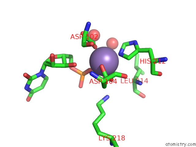
Manganese Binding Sites:
The binding sites of Manganese atom in the Crystal Structure of Human Glycogenin-1 (GYG1) T83M Mutant Complexed with Manganese and Udp
(pdb code 3rmv). This binding sites where shown within
5.0 Angstroms radius around Manganese atom.
In total only one binding site of Manganese was determined in the Crystal Structure of Human Glycogenin-1 (GYG1) T83M Mutant Complexed with Manganese and Udp, PDB code: 3rmv:
In total only one binding site of Manganese was determined in the Crystal Structure of Human Glycogenin-1 (GYG1) T83M Mutant Complexed with Manganese and Udp, PDB code: 3rmv:
Manganese binding site 1 out of 1 in 3rmv
Go back to
Manganese binding site 1 out
of 1 in the Crystal Structure of Human Glycogenin-1 (GYG1) T83M Mutant Complexed with Manganese and Udp
Mono view

Stereo pair view
Mono view
Stereo pair view
A full contact list of Manganese with other atoms in the Mn binding
site number 1 of Crystal Structure of Human Glycogenin-1 (GYG1) T83M Mutant Complexed with Manganese and Udp within 5.0Å range:
|
Reference:
A.Chaikuad,
D.S.Froese,
G.Berridge,
F.Von Delft,
U.Oppermann,
W.W.Yue.
Conformational Plasticity of Glycogenin and Its Maltosaccharide Substrate During Glycogen Biogenesis. Proc.Natl.Acad.Sci.Usa V. 108 21028 2011.
ISSN: ISSN 0027-8424
PubMed: 22160680
DOI: 10.1073/PNAS.1113921108
Page generated: Sat Oct 5 17:50:06 2024
ISSN: ISSN 0027-8424
PubMed: 22160680
DOI: 10.1073/PNAS.1113921108
Last articles
Cl in 7VUJCl in 7VUX
Cl in 7VUE
Cl in 7VUI
Cl in 7VUH
Cl in 7VUG
Cl in 7VU6
Cl in 7VS8
Cl in 7VTX
Cl in 7VTW