Manganese »
PDB 3q7c-3rl4 »
3ras »
Manganese in PDB 3ras: Crystal Structure of 1-Deoxy-D-Xylulose 5-Phosphate Reductoisomerase (Dxr) Complexed with A Lipophilic Phosphonate Inhibitor
Enzymatic activity of Crystal Structure of 1-Deoxy-D-Xylulose 5-Phosphate Reductoisomerase (Dxr) Complexed with A Lipophilic Phosphonate Inhibitor
All present enzymatic activity of Crystal Structure of 1-Deoxy-D-Xylulose 5-Phosphate Reductoisomerase (Dxr) Complexed with A Lipophilic Phosphonate Inhibitor:
1.1.1.267;
1.1.1.267;
Protein crystallography data
The structure of Crystal Structure of 1-Deoxy-D-Xylulose 5-Phosphate Reductoisomerase (Dxr) Complexed with A Lipophilic Phosphonate Inhibitor, PDB code: 3ras
was solved by
J.Diao,
L.Deng,
B.V.V.Prasad,
Y.Song,
with X-Ray Crystallography technique. A brief refinement statistics is given in the table below:
| Resolution Low / High (Å) | 33.45 / 2.55 |
| Space group | P 1 21 1 |
| Cell size a, b, c (Å), α, β, γ (°) | 67.250, 65.179, 85.408, 90.00, 101.28, 90.00 |
| R / Rfree (%) | 20.8 / 27.8 |
Other elements in 3ras:
The structure of Crystal Structure of 1-Deoxy-D-Xylulose 5-Phosphate Reductoisomerase (Dxr) Complexed with A Lipophilic Phosphonate Inhibitor also contains other interesting chemical elements:
| Chlorine | (Cl) | 8 atoms |
Manganese Binding Sites:
The binding sites of Manganese atom in the Crystal Structure of 1-Deoxy-D-Xylulose 5-Phosphate Reductoisomerase (Dxr) Complexed with A Lipophilic Phosphonate Inhibitor
(pdb code 3ras). This binding sites where shown within
5.0 Angstroms radius around Manganese atom.
In total 2 binding sites of Manganese where determined in the Crystal Structure of 1-Deoxy-D-Xylulose 5-Phosphate Reductoisomerase (Dxr) Complexed with A Lipophilic Phosphonate Inhibitor, PDB code: 3ras:
Jump to Manganese binding site number: 1; 2;
In total 2 binding sites of Manganese where determined in the Crystal Structure of 1-Deoxy-D-Xylulose 5-Phosphate Reductoisomerase (Dxr) Complexed with A Lipophilic Phosphonate Inhibitor, PDB code: 3ras:
Jump to Manganese binding site number: 1; 2;
Manganese binding site 1 out of 2 in 3ras
Go back to
Manganese binding site 1 out
of 2 in the Crystal Structure of 1-Deoxy-D-Xylulose 5-Phosphate Reductoisomerase (Dxr) Complexed with A Lipophilic Phosphonate Inhibitor

Mono view
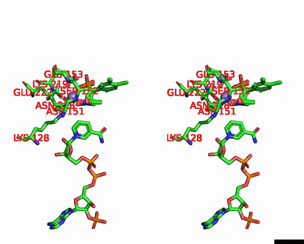
Stereo pair view
Mono view
Stereo pair view
A full contact list of Manganese with other atoms in the Mn binding
site number 1 of Crystal Structure of 1-Deoxy-D-Xylulose 5-Phosphate Reductoisomerase (Dxr) Complexed with A Lipophilic Phosphonate Inhibitor within 5.0Å range:
|
Manganese binding site 2 out of 2 in 3ras
Go back to
Manganese binding site 2 out
of 2 in the Crystal Structure of 1-Deoxy-D-Xylulose 5-Phosphate Reductoisomerase (Dxr) Complexed with A Lipophilic Phosphonate Inhibitor
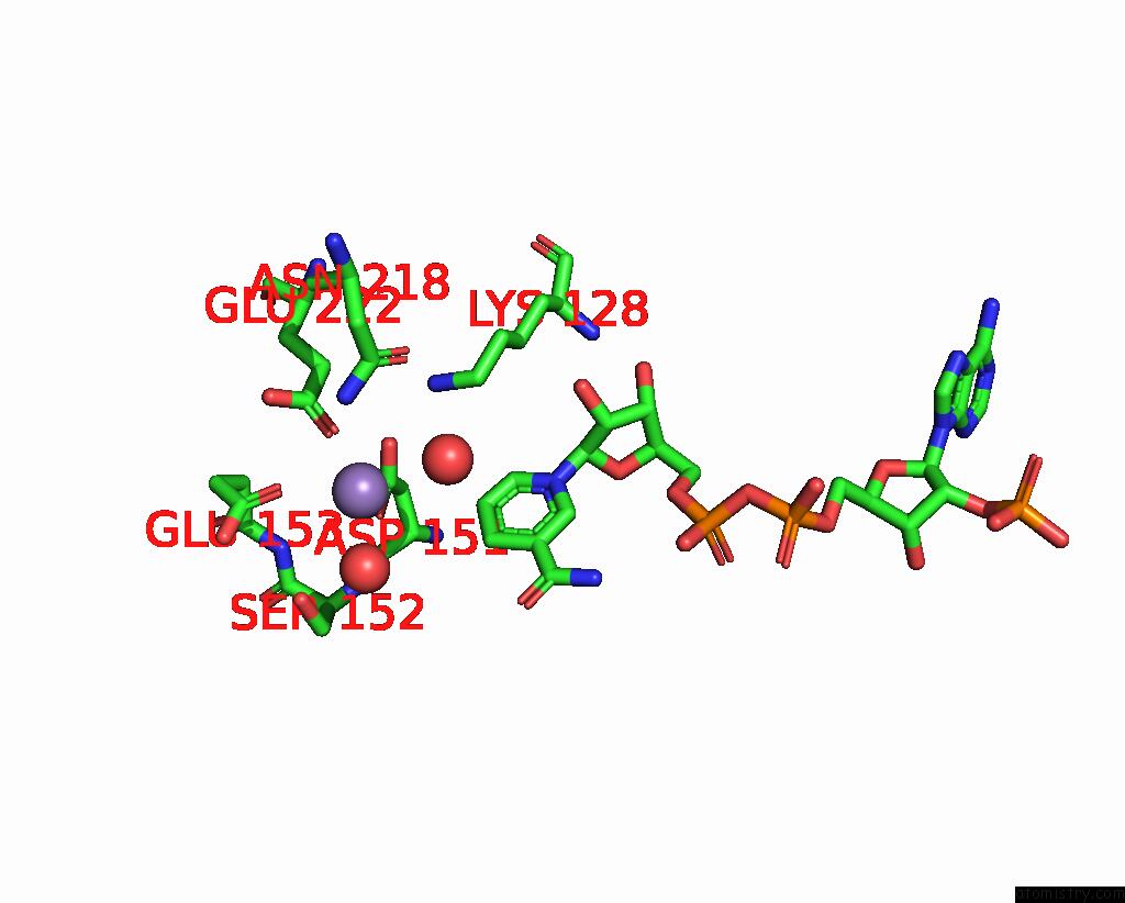
Mono view

Stereo pair view
Mono view
Stereo pair view
A full contact list of Manganese with other atoms in the Mn binding
site number 2 of Crystal Structure of 1-Deoxy-D-Xylulose 5-Phosphate Reductoisomerase (Dxr) Complexed with A Lipophilic Phosphonate Inhibitor within 5.0Å range:
|
Reference:
L.Deng,
J.Diao,
P.Chen,
V.Pujari,
Y.Yao,
G.Cheng,
D.C.Crick,
B.V.Prasad,
Y.Song.
Inhibition of 1-Deoxy-D-Xylulose-5-Phosphate Reductoisomerase By Lipophilic Phosphonates: Sar, Qsar, and Crystallographic Studies. J.Med.Chem. V. 54 4721 2011.
ISSN: ISSN 0022-2623
PubMed: 21561155
DOI: 10.1021/JM200363D
Page generated: Sat Oct 5 17:43:54 2024
ISSN: ISSN 0022-2623
PubMed: 21561155
DOI: 10.1021/JM200363D
Last articles
Fe in 2YXOFe in 2YRS
Fe in 2YXC
Fe in 2YNM
Fe in 2YVJ
Fe in 2YP1
Fe in 2YU2
Fe in 2YU1
Fe in 2YQB
Fe in 2YOO