Manganese »
PDB 3q7c-3rl4 »
3ql3 »
Manganese in PDB 3ql3: Re-Refined Coordinates For Pdb Entry 1RX2
Enzymatic activity of Re-Refined Coordinates For Pdb Entry 1RX2
All present enzymatic activity of Re-Refined Coordinates For Pdb Entry 1RX2:
1.5.1.3;
1.5.1.3;
Protein crystallography data
The structure of Re-Refined Coordinates For Pdb Entry 1RX2, PDB code: 3ql3
was solved by
G.Bhabha,
D.C.Ekiert,
P.E.Wright,
I.A.Wilson,
with X-Ray Crystallography technique. A brief refinement statistics is given in the table below:
| Resolution Low / High (Å) | 41.00 / 1.80 |
| Space group | P 21 21 21 |
| Cell size a, b, c (Å), α, β, γ (°) | 34.320, 45.510, 98.910, 90.00, 90.00, 90.00 |
| R / Rfree (%) | 17.7 / 21.3 |
Manganese Binding Sites:
The binding sites of Manganese atom in the Re-Refined Coordinates For Pdb Entry 1RX2
(pdb code 3ql3). This binding sites where shown within
5.0 Angstroms radius around Manganese atom.
In total 2 binding sites of Manganese where determined in the Re-Refined Coordinates For Pdb Entry 1RX2, PDB code: 3ql3:
Jump to Manganese binding site number: 1; 2;
In total 2 binding sites of Manganese where determined in the Re-Refined Coordinates For Pdb Entry 1RX2, PDB code: 3ql3:
Jump to Manganese binding site number: 1; 2;
Manganese binding site 1 out of 2 in 3ql3
Go back to
Manganese binding site 1 out
of 2 in the Re-Refined Coordinates For Pdb Entry 1RX2
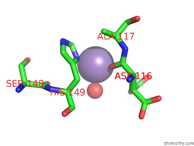
Mono view
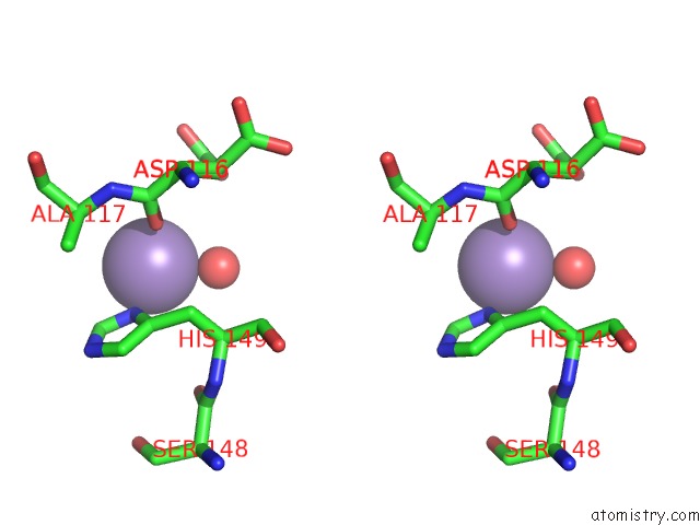
Stereo pair view
Mono view
Stereo pair view
A full contact list of Manganese with other atoms in the Mn binding
site number 1 of Re-Refined Coordinates For Pdb Entry 1RX2 within 5.0Å range:
|
Manganese binding site 2 out of 2 in 3ql3
Go back to
Manganese binding site 2 out
of 2 in the Re-Refined Coordinates For Pdb Entry 1RX2
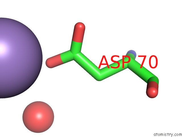
Mono view
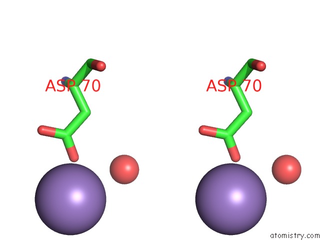
Stereo pair view
Mono view
Stereo pair view
A full contact list of Manganese with other atoms in the Mn binding
site number 2 of Re-Refined Coordinates For Pdb Entry 1RX2 within 5.0Å range:
|
Reference:
G.Bhabha,
J.Lee,
D.C.Ekiert,
J.Gam,
I.A.Wilson,
H.J.Dyson,
S.J.Benkovic,
P.E.Wright.
A Dynamic Knockout Reveals That Conformational Fluctuations Influence the Chemical Step of Enzyme Catalysis. Science V. 332 234 2011.
ISSN: ISSN 0036-8075
PubMed: 21474759
DOI: 10.1126/SCIENCE.1198542
Page generated: Sat Oct 5 17:41:30 2024
ISSN: ISSN 0036-8075
PubMed: 21474759
DOI: 10.1126/SCIENCE.1198542
Last articles
Zn in 9JYWZn in 9IR4
Zn in 9IR3
Zn in 9GMX
Zn in 9GMW
Zn in 9JEJ
Zn in 9ERF
Zn in 9ERE
Zn in 9EGV
Zn in 9EGW