Manganese »
PDB 3q7c-3rl4 »
3qin »
Manganese in PDB 3qin: Crystal Structure of Hiv-1 Rnase H P15 with Engineered E. Coli Loop and Pyrimidinol Carboxylic Acid Inhibitor
Enzymatic activity of Crystal Structure of Hiv-1 Rnase H P15 with Engineered E. Coli Loop and Pyrimidinol Carboxylic Acid Inhibitor
All present enzymatic activity of Crystal Structure of Hiv-1 Rnase H P15 with Engineered E. Coli Loop and Pyrimidinol Carboxylic Acid Inhibitor:
2.7.7.49; 2.7.7.7; 3.1.13.2; 3.1.26.13; 3.4.23.16;
2.7.7.49; 2.7.7.7; 3.1.13.2; 3.1.26.13; 3.4.23.16;
Protein crystallography data
The structure of Crystal Structure of Hiv-1 Rnase H P15 with Engineered E. Coli Loop and Pyrimidinol Carboxylic Acid Inhibitor, PDB code: 3qin
was solved by
E.B.Lansdon,
T.A.Kirschberg,
with X-Ray Crystallography technique. A brief refinement statistics is given in the table below:
| Resolution Low / High (Å) | 23.12 / 1.70 |
| Space group | I 21 21 21 |
| Cell size a, b, c (Å), α, β, γ (°) | 38.841, 90.370, 111.922, 90.00, 90.00, 90.00 |
| R / Rfree (%) | 22 / 25.3 |
Other elements in 3qin:
The structure of Crystal Structure of Hiv-1 Rnase H P15 with Engineered E. Coli Loop and Pyrimidinol Carboxylic Acid Inhibitor also contains other interesting chemical elements:
| Bromine | (Br) | 1 atom |
Manganese Binding Sites:
The binding sites of Manganese atom in the Crystal Structure of Hiv-1 Rnase H P15 with Engineered E. Coli Loop and Pyrimidinol Carboxylic Acid Inhibitor
(pdb code 3qin). This binding sites where shown within
5.0 Angstroms radius around Manganese atom.
In total 2 binding sites of Manganese where determined in the Crystal Structure of Hiv-1 Rnase H P15 with Engineered E. Coli Loop and Pyrimidinol Carboxylic Acid Inhibitor, PDB code: 3qin:
Jump to Manganese binding site number: 1; 2;
In total 2 binding sites of Manganese where determined in the Crystal Structure of Hiv-1 Rnase H P15 with Engineered E. Coli Loop and Pyrimidinol Carboxylic Acid Inhibitor, PDB code: 3qin:
Jump to Manganese binding site number: 1; 2;
Manganese binding site 1 out of 2 in 3qin
Go back to
Manganese binding site 1 out
of 2 in the Crystal Structure of Hiv-1 Rnase H P15 with Engineered E. Coli Loop and Pyrimidinol Carboxylic Acid Inhibitor
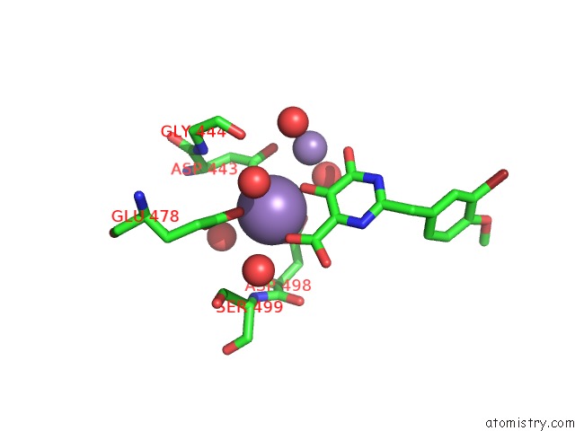
Mono view
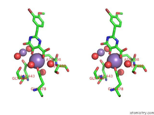
Stereo pair view
Mono view
Stereo pair view
A full contact list of Manganese with other atoms in the Mn binding
site number 1 of Crystal Structure of Hiv-1 Rnase H P15 with Engineered E. Coli Loop and Pyrimidinol Carboxylic Acid Inhibitor within 5.0Å range:
|
Manganese binding site 2 out of 2 in 3qin
Go back to
Manganese binding site 2 out
of 2 in the Crystal Structure of Hiv-1 Rnase H P15 with Engineered E. Coli Loop and Pyrimidinol Carboxylic Acid Inhibitor
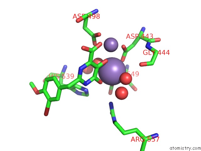
Mono view
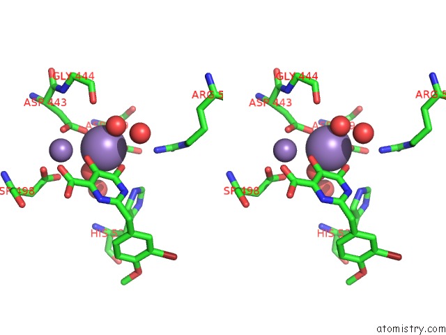
Stereo pair view
Mono view
Stereo pair view
A full contact list of Manganese with other atoms in the Mn binding
site number 2 of Crystal Structure of Hiv-1 Rnase H P15 with Engineered E. Coli Loop and Pyrimidinol Carboxylic Acid Inhibitor within 5.0Å range:
|
Reference:
E.B.Lansdon,
Q.Liu,
S.A.Leavitt,
M.Balakrishnan,
J.K.Perry,
C.Lancaster-Moyer,
N.Kutty,
X.Liu,
N.H.Squires,
W.J.Watkins,
T.A.Kirschberg.
Structural and Binding Analysis of Pyrimidinol Carboxylic Acid and N-Hydroxy Quinazolinedione Hiv-1 Rnase H Inhibitors. Antimicrob.Agents Chemother. V. 55 2905 2011.
ISSN: ISSN 0066-4804
PubMed: 21464257
DOI: 10.1128/AAC.01594-10
Page generated: Sat Oct 5 17:40:49 2024
ISSN: ISSN 0066-4804
PubMed: 21464257
DOI: 10.1128/AAC.01594-10
Last articles
Zn in 9JYWZn in 9IR4
Zn in 9IR3
Zn in 9GMX
Zn in 9GMW
Zn in 9JEJ
Zn in 9ERF
Zn in 9ERE
Zn in 9EGV
Zn in 9EGW