Manganese »
PDB 3q7c-3rl4 »
3qh8 »
Manganese in PDB 3qh8: Crystal Structure of A Beta-Lactamase-Like Protein Bound to Amp From Brucella Melitensis, Long Wavelength Synchrotron Data
Protein crystallography data
The structure of Crystal Structure of A Beta-Lactamase-Like Protein Bound to Amp From Brucella Melitensis, Long Wavelength Synchrotron Data, PDB code: 3qh8
was solved by
Seattle Structural Genomics Center For Infectious Disease (Ssgcid),
with X-Ray Crystallography technique. A brief refinement statistics is given in the table below:
| Resolution Low / High (Å) | 18.88 / 1.60 |
| Space group | C 2 2 21 |
| Cell size a, b, c (Å), α, β, γ (°) | 73.210, 75.530, 98.940, 90.00, 90.00, 90.00 |
| R / Rfree (%) | 14.3 / 16.6 |
Other elements in 3qh8:
The structure of Crystal Structure of A Beta-Lactamase-Like Protein Bound to Amp From Brucella Melitensis, Long Wavelength Synchrotron Data also contains other interesting chemical elements:
| Potassium | (K) | 1 atom |
Manganese Binding Sites:
The binding sites of Manganese atom in the Crystal Structure of A Beta-Lactamase-Like Protein Bound to Amp From Brucella Melitensis, Long Wavelength Synchrotron Data
(pdb code 3qh8). This binding sites where shown within
5.0 Angstroms radius around Manganese atom.
In total 2 binding sites of Manganese where determined in the Crystal Structure of A Beta-Lactamase-Like Protein Bound to Amp From Brucella Melitensis, Long Wavelength Synchrotron Data, PDB code: 3qh8:
Jump to Manganese binding site number: 1; 2;
In total 2 binding sites of Manganese where determined in the Crystal Structure of A Beta-Lactamase-Like Protein Bound to Amp From Brucella Melitensis, Long Wavelength Synchrotron Data, PDB code: 3qh8:
Jump to Manganese binding site number: 1; 2;
Manganese binding site 1 out of 2 in 3qh8
Go back to
Manganese binding site 1 out
of 2 in the Crystal Structure of A Beta-Lactamase-Like Protein Bound to Amp From Brucella Melitensis, Long Wavelength Synchrotron Data
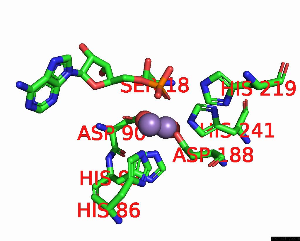
Mono view
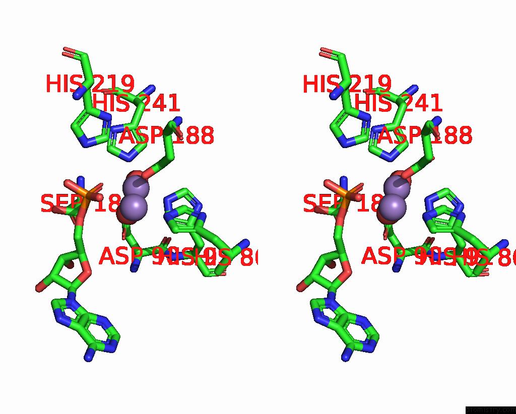
Stereo pair view
Mono view
Stereo pair view
A full contact list of Manganese with other atoms in the Mn binding
site number 1 of Crystal Structure of A Beta-Lactamase-Like Protein Bound to Amp From Brucella Melitensis, Long Wavelength Synchrotron Data within 5.0Å range:
|
Manganese binding site 2 out of 2 in 3qh8
Go back to
Manganese binding site 2 out
of 2 in the Crystal Structure of A Beta-Lactamase-Like Protein Bound to Amp From Brucella Melitensis, Long Wavelength Synchrotron Data
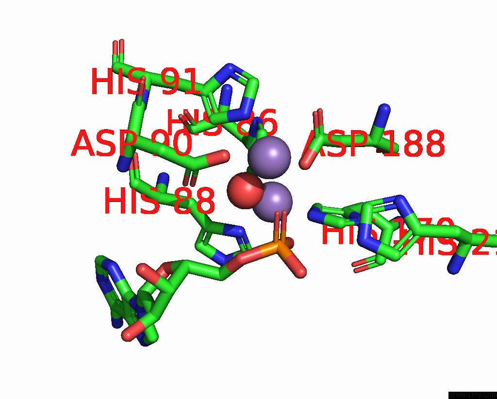
Mono view
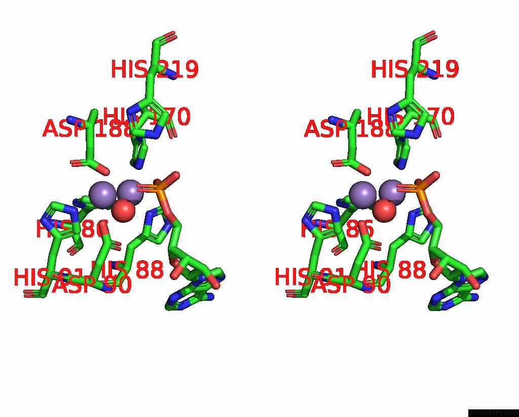
Stereo pair view
Mono view
Stereo pair view
A full contact list of Manganese with other atoms in the Mn binding
site number 2 of Crystal Structure of A Beta-Lactamase-Like Protein Bound to Amp From Brucella Melitensis, Long Wavelength Synchrotron Data within 5.0Å range:
|
Reference:
J.Abendroth,
B.Sankaran,
T.E.Edwards,
A.S.Gardberg,
S.Dieterich,
J.Bhandari,
A.J.Napuli,
W.C.Van Voorhis,
B.L.Staker,
P.J.Myler,
L.J.Stewart.
Crystal Structure of A Beta-Lactamase-Like Protein Bound to Amp From Brucella Melitensis, Long Wavelength Synchrotron Data Acta Crystallogr.,Sect.F V. 67 1106 2011.
ISSN: ESSN 1744-3091
PubMed: 21904058
DOI: 10.1107/S1744309111010220
Page generated: Sat Oct 5 17:40:46 2024
ISSN: ESSN 1744-3091
PubMed: 21904058
DOI: 10.1107/S1744309111010220
Last articles
Fe in 2YXOFe in 2YRS
Fe in 2YXC
Fe in 2YNM
Fe in 2YVJ
Fe in 2YP1
Fe in 2YU2
Fe in 2YU1
Fe in 2YQB
Fe in 2YOO