Manganese »
PDB 3q7c-3rl4 »
3qfo »
Manganese in PDB 3qfo: Crystal Structure of Streptococcal Asymmetric AP4A Hydrolase and Phosphodiesterase SPR1479/Saph Im Complex with Amp
Protein crystallography data
The structure of Crystal Structure of Streptococcal Asymmetric AP4A Hydrolase and Phosphodiesterase SPR1479/Saph Im Complex with Amp, PDB code: 3qfo
was solved by
Y.L.Jiang,
J.W.Zhang,
W.L.Yu,
W.Cheng,
C.C.Zhang,
C.Z.Zhou,
Y.Chen,
with X-Ray Crystallography technique. A brief refinement statistics is given in the table below:
| Resolution Low / High (Å) | 50.00 / 2.20 |
| Space group | P 21 21 21 |
| Cell size a, b, c (Å), α, β, γ (°) | 57.654, 74.940, 149.130, 90.00, 90.00, 90.00 |
| R / Rfree (%) | 18.5 / 24.5 |
Other elements in 3qfo:
The structure of Crystal Structure of Streptococcal Asymmetric AP4A Hydrolase and Phosphodiesterase SPR1479/Saph Im Complex with Amp also contains other interesting chemical elements:
| Iron | (Fe) | 2 atoms |
Manganese Binding Sites:
The binding sites of Manganese atom in the Crystal Structure of Streptococcal Asymmetric AP4A Hydrolase and Phosphodiesterase SPR1479/Saph Im Complex with Amp
(pdb code 3qfo). This binding sites where shown within
5.0 Angstroms radius around Manganese atom.
In total 2 binding sites of Manganese where determined in the Crystal Structure of Streptococcal Asymmetric AP4A Hydrolase and Phosphodiesterase SPR1479/Saph Im Complex with Amp, PDB code: 3qfo:
Jump to Manganese binding site number: 1; 2;
In total 2 binding sites of Manganese where determined in the Crystal Structure of Streptococcal Asymmetric AP4A Hydrolase and Phosphodiesterase SPR1479/Saph Im Complex with Amp, PDB code: 3qfo:
Jump to Manganese binding site number: 1; 2;
Manganese binding site 1 out of 2 in 3qfo
Go back to
Manganese binding site 1 out
of 2 in the Crystal Structure of Streptococcal Asymmetric AP4A Hydrolase and Phosphodiesterase SPR1479/Saph Im Complex with Amp
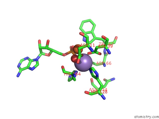
Mono view
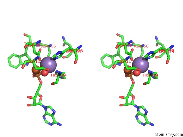
Stereo pair view
Mono view
Stereo pair view
A full contact list of Manganese with other atoms in the Mn binding
site number 1 of Crystal Structure of Streptococcal Asymmetric AP4A Hydrolase and Phosphodiesterase SPR1479/Saph Im Complex with Amp within 5.0Å range:
|
Manganese binding site 2 out of 2 in 3qfo
Go back to
Manganese binding site 2 out
of 2 in the Crystal Structure of Streptococcal Asymmetric AP4A Hydrolase and Phosphodiesterase SPR1479/Saph Im Complex with Amp
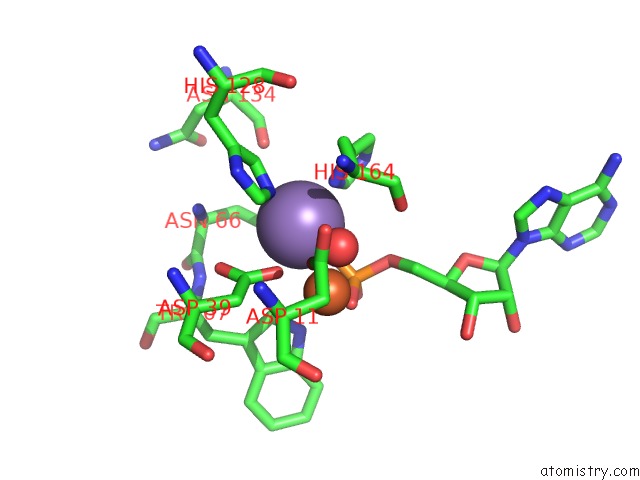
Mono view
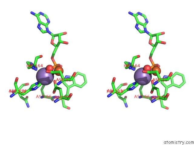
Stereo pair view
Mono view
Stereo pair view
A full contact list of Manganese with other atoms in the Mn binding
site number 2 of Crystal Structure of Streptococcal Asymmetric AP4A Hydrolase and Phosphodiesterase SPR1479/Saph Im Complex with Amp within 5.0Å range:
|
Reference:
Y.L.Jiang,
J.W.Zhang,
W.L.Yu,
W.Cheng,
C.C.Zhang,
C.Frolet,
A.-M.Di-Guilmi,
T.Vernet,
C.Z.Zhou,
Y.Chen.
Structural and Enzymatic Characterization of A Streptococcal Atp/Diadenosine Polyphosphate and Phosphodiester Hydrolase SPR1479/Saph To Be Published.
Page generated: Sat Oct 5 17:40:06 2024
Last articles
Zn in 9J0NZn in 9J0O
Zn in 9J0P
Zn in 9FJX
Zn in 9EKB
Zn in 9C0F
Zn in 9CAH
Zn in 9CH0
Zn in 9CH3
Zn in 9CH1