Manganese »
PDB 3ot9-3q4q »
3q2v »
Manganese in PDB 3q2v: Crystal Structure of Mouse E-Cadherin Ectodomain
Protein crystallography data
The structure of Crystal Structure of Mouse E-Cadherin Ectodomain, PDB code: 3q2v
was solved by
X.Jin,
O.J.Harrison,
L.Shapiro,
with X-Ray Crystallography technique. A brief refinement statistics is given in the table below:
| Resolution Low / High (Å) | 19.92 / 3.40 |
| Space group | C 1 2 1 |
| Cell size a, b, c (Å), α, β, γ (°) | 119.140, 79.697, 176.001, 90.00, 98.56, 90.00 |
| R / Rfree (%) | 23 / 29.3 |
Other elements in 3q2v:
The structure of Crystal Structure of Mouse E-Cadherin Ectodomain also contains other interesting chemical elements:
| Calcium | (Ca) | 24 atoms |
Manganese Binding Sites:
The binding sites of Manganese atom in the Crystal Structure of Mouse E-Cadherin Ectodomain
(pdb code 3q2v). This binding sites where shown within
5.0 Angstroms radius around Manganese atom.
In total 2 binding sites of Manganese where determined in the Crystal Structure of Mouse E-Cadherin Ectodomain, PDB code: 3q2v:
Jump to Manganese binding site number: 1; 2;
In total 2 binding sites of Manganese where determined in the Crystal Structure of Mouse E-Cadherin Ectodomain, PDB code: 3q2v:
Jump to Manganese binding site number: 1; 2;
Manganese binding site 1 out of 2 in 3q2v
Go back to
Manganese binding site 1 out
of 2 in the Crystal Structure of Mouse E-Cadherin Ectodomain
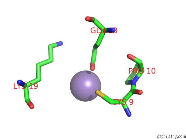
Mono view
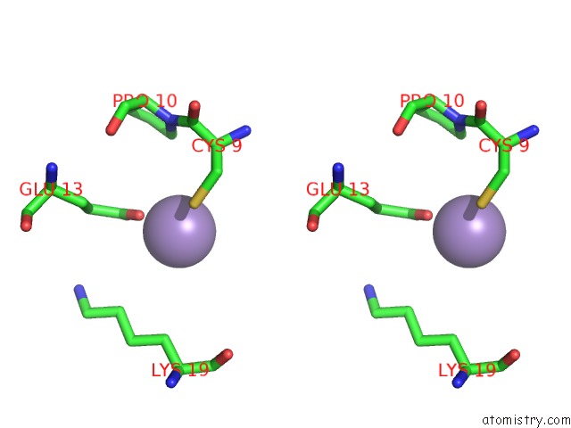
Stereo pair view
Mono view
Stereo pair view
A full contact list of Manganese with other atoms in the Mn binding
site number 1 of Crystal Structure of Mouse E-Cadherin Ectodomain within 5.0Å range:
|
Manganese binding site 2 out of 2 in 3q2v
Go back to
Manganese binding site 2 out
of 2 in the Crystal Structure of Mouse E-Cadherin Ectodomain
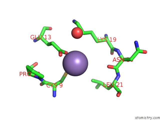
Mono view
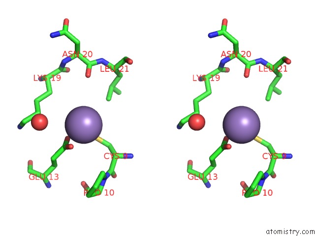
Stereo pair view
Mono view
Stereo pair view
A full contact list of Manganese with other atoms in the Mn binding
site number 2 of Crystal Structure of Mouse E-Cadherin Ectodomain within 5.0Å range:
|
Reference:
O.J.Harrison,
X.Jin,
S.Hong,
F.Bahna,
G.Ahlsen,
J.Brasch,
Y.Wu,
J.Vendome,
K.Felsovalyi,
C.M.Hampton,
R.B.Troyanovsky,
A.Ben-Shaul,
J.Frank,
S.M.Troyanovsky,
L.Shapiro,
B.Honig.
The Extracellular Architecture of Adherens Junctions Revealed By Crystal Structures of Type I Cadherins. Structure V. 19 244 2011.
ISSN: ISSN 0969-2126
PubMed: 21300292
DOI: 10.1016/J.STR.2010.11.016
Page generated: Sat Oct 5 17:35:38 2024
ISSN: ISSN 0969-2126
PubMed: 21300292
DOI: 10.1016/J.STR.2010.11.016
Last articles
Zn in 9J0NZn in 9J0O
Zn in 9J0P
Zn in 9FJX
Zn in 9EKB
Zn in 9C0F
Zn in 9CAH
Zn in 9CH0
Zn in 9CH3
Zn in 9CH1