Manganese »
PDB 3ot9-3q4q »
3pzt »
Manganese in PDB 3pzt: Structure of the Endo-1,4-Beta-Glucanase From Bacillus Subtilis 168 with Manganese(II) Ion
Enzymatic activity of Structure of the Endo-1,4-Beta-Glucanase From Bacillus Subtilis 168 with Manganese(II) Ion
All present enzymatic activity of Structure of the Endo-1,4-Beta-Glucanase From Bacillus Subtilis 168 with Manganese(II) Ion:
3.2.1.4;
3.2.1.4;
Protein crystallography data
The structure of Structure of the Endo-1,4-Beta-Glucanase From Bacillus Subtilis 168 with Manganese(II) Ion, PDB code: 3pzt
was solved by
C.R.Santos,
J.H.Paiva,
P.K.Akao,
A.N.Meza,
J.C.Silva,
F.M.Squina,
R.J.Ward,
R.Ruller,
M.T.Murakami,
with X-Ray Crystallography technique. A brief refinement statistics is given in the table below:
| Resolution Low / High (Å) | 42.40 / 1.97 |
| Space group | P 21 21 21 |
| Cell size a, b, c (Å), α, β, γ (°) | 49.526, 110.617, 122.262, 90.00, 90.00, 90.00 |
| R / Rfree (%) | 16.4 / 20.6 |
Manganese Binding Sites:
The binding sites of Manganese atom in the Structure of the Endo-1,4-Beta-Glucanase From Bacillus Subtilis 168 with Manganese(II) Ion
(pdb code 3pzt). This binding sites where shown within
5.0 Angstroms radius around Manganese atom.
In total 2 binding sites of Manganese where determined in the Structure of the Endo-1,4-Beta-Glucanase From Bacillus Subtilis 168 with Manganese(II) Ion, PDB code: 3pzt:
Jump to Manganese binding site number: 1; 2;
In total 2 binding sites of Manganese where determined in the Structure of the Endo-1,4-Beta-Glucanase From Bacillus Subtilis 168 with Manganese(II) Ion, PDB code: 3pzt:
Jump to Manganese binding site number: 1; 2;
Manganese binding site 1 out of 2 in 3pzt
Go back to
Manganese binding site 1 out
of 2 in the Structure of the Endo-1,4-Beta-Glucanase From Bacillus Subtilis 168 with Manganese(II) Ion
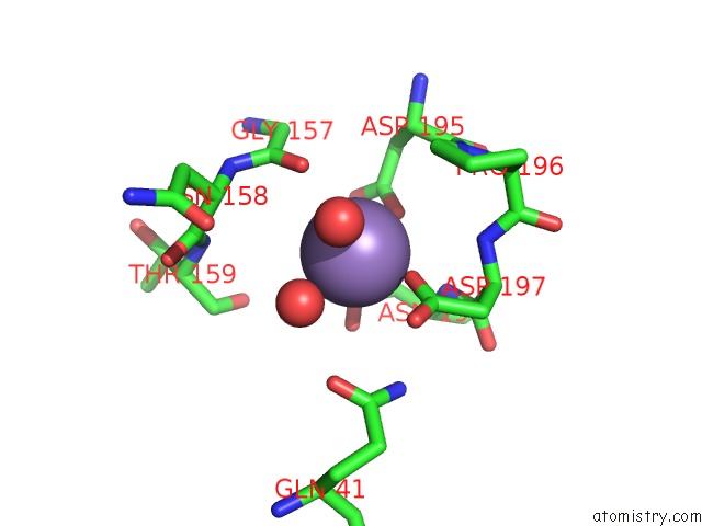
Mono view
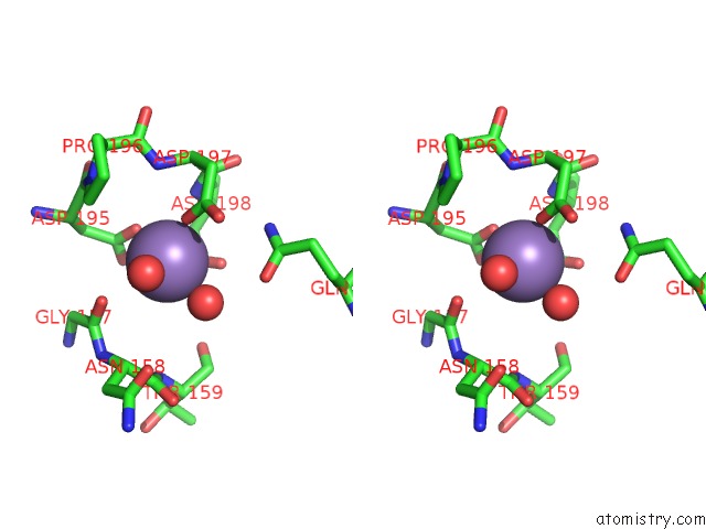
Stereo pair view
Mono view
Stereo pair view
A full contact list of Manganese with other atoms in the Mn binding
site number 1 of Structure of the Endo-1,4-Beta-Glucanase From Bacillus Subtilis 168 with Manganese(II) Ion within 5.0Å range:
|
Manganese binding site 2 out of 2 in 3pzt
Go back to
Manganese binding site 2 out
of 2 in the Structure of the Endo-1,4-Beta-Glucanase From Bacillus Subtilis 168 with Manganese(II) Ion
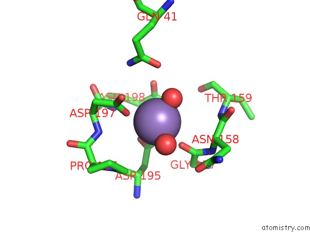
Mono view
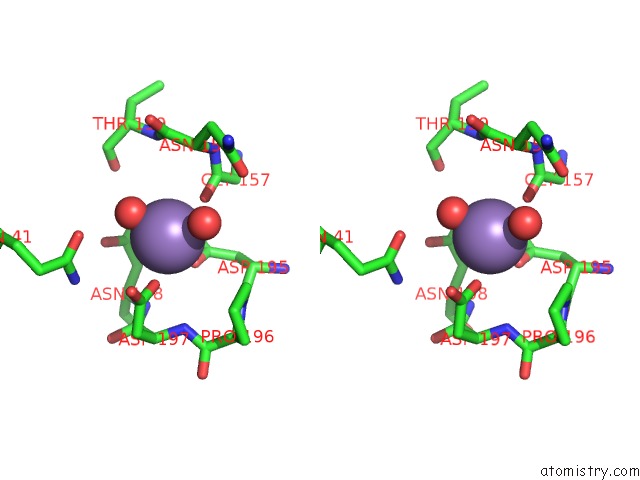
Stereo pair view
Mono view
Stereo pair view
A full contact list of Manganese with other atoms in the Mn binding
site number 2 of Structure of the Endo-1,4-Beta-Glucanase From Bacillus Subtilis 168 with Manganese(II) Ion within 5.0Å range:
|
Reference:
C.R.Santos,
J.H.Paiva,
M.L.Sforca,
J.L.Neves,
R.Z.Navarro,
J.Cota,
P.K.Akao,
Z.B.Hoffmam,
A.N.Meza,
J.H.Smetana,
M.L.Nogueira,
I.Polikarpov,
J.Xavier-Neto,
F.M.Squina,
R.J.Ward,
R.Ruller,
A.C.Zeri,
M.T.Murakami.
Dissecting Structure-Function-Stability Relationships of A Thermostable GH5-CBM3 Cellulase From Bacillus Subtilis 168. Biochem.J. V. 441 95 2012.
ISSN: ISSN 0264-6021
PubMed: 21880019
DOI: 10.1042/BJ20110869
Page generated: Sat Oct 5 17:35:24 2024
ISSN: ISSN 0264-6021
PubMed: 21880019
DOI: 10.1042/BJ20110869
Last articles
Zn in 9J0NZn in 9J0O
Zn in 9J0P
Zn in 9FJX
Zn in 9EKB
Zn in 9C0F
Zn in 9CAH
Zn in 9CH0
Zn in 9CH3
Zn in 9CH1