Manganese »
PDB 3n39-3orm »
3n3a »
Manganese in PDB 3n3a: Ribonucleotide Reductase Dimanganese(II)-Nrdf From Escherichia Coli in Complex with Reduced Nrdi
Enzymatic activity of Ribonucleotide Reductase Dimanganese(II)-Nrdf From Escherichia Coli in Complex with Reduced Nrdi
All present enzymatic activity of Ribonucleotide Reductase Dimanganese(II)-Nrdf From Escherichia Coli in Complex with Reduced Nrdi:
1.17.4.1;
1.17.4.1;
Protein crystallography data
The structure of Ribonucleotide Reductase Dimanganese(II)-Nrdf From Escherichia Coli in Complex with Reduced Nrdi, PDB code: 3n3a
was solved by
A.K.Boal,
J.A.Cotruvo Jr.,
J.Stubbe,
A.C.Rosenzweig,
with X-Ray Crystallography technique. A brief refinement statistics is given in the table below:
| Resolution Low / High (Å) | 29.58 / 1.99 |
| Space group | P 21 21 21 |
| Cell size a, b, c (Å), α, β, γ (°) | 74.863, 90.678, 143.837, 90.00, 90.00, 90.00 |
| R / Rfree (%) | 20.3 / 22.7 |
Manganese Binding Sites:
The binding sites of Manganese atom in the Ribonucleotide Reductase Dimanganese(II)-Nrdf From Escherichia Coli in Complex with Reduced Nrdi
(pdb code 3n3a). This binding sites where shown within
5.0 Angstroms radius around Manganese atom.
In total 4 binding sites of Manganese where determined in the Ribonucleotide Reductase Dimanganese(II)-Nrdf From Escherichia Coli in Complex with Reduced Nrdi, PDB code: 3n3a:
Jump to Manganese binding site number: 1; 2; 3; 4;
In total 4 binding sites of Manganese where determined in the Ribonucleotide Reductase Dimanganese(II)-Nrdf From Escherichia Coli in Complex with Reduced Nrdi, PDB code: 3n3a:
Jump to Manganese binding site number: 1; 2; 3; 4;
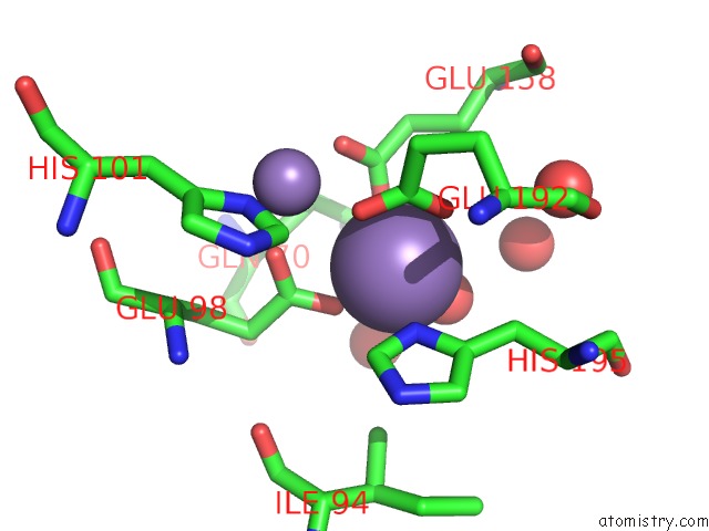
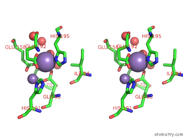
Manganese binding site 1 out of 4 in 3n3a
Go back to
Manganese binding site 1 out
of 4 in the Ribonucleotide Reductase Dimanganese(II)-Nrdf From Escherichia Coli in Complex with Reduced Nrdi
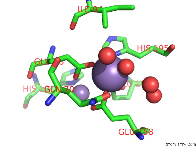
Mono view
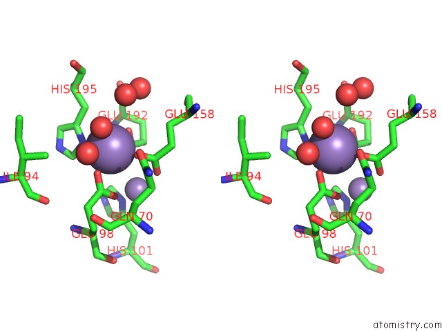
Stereo pair view
Mono view
Stereo pair view
A full contact list of Manganese with other atoms in the Mn binding
site number 1 of Ribonucleotide Reductase Dimanganese(II)-Nrdf From Escherichia Coli in Complex with Reduced Nrdi within 5.0Å range:
|
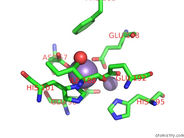
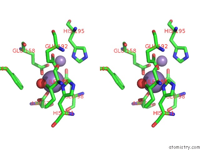
Manganese binding site 2 out of 4 in 3n3a
Go back to
Manganese binding site 2 out
of 4 in the Ribonucleotide Reductase Dimanganese(II)-Nrdf From Escherichia Coli in Complex with Reduced Nrdi
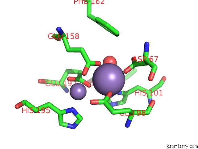
Mono view
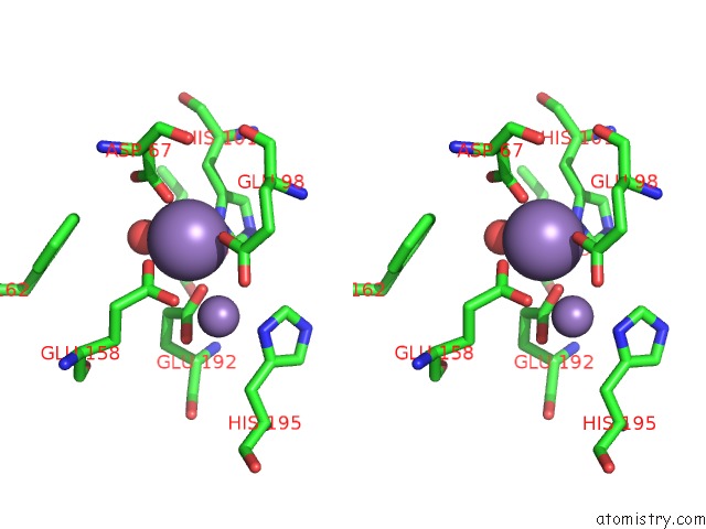
Stereo pair view
Mono view
Stereo pair view
A full contact list of Manganese with other atoms in the Mn binding
site number 2 of Ribonucleotide Reductase Dimanganese(II)-Nrdf From Escherichia Coli in Complex with Reduced Nrdi within 5.0Å range:
|
Manganese binding site 3 out of 4 in 3n3a
Go back to
Manganese binding site 3 out
of 4 in the Ribonucleotide Reductase Dimanganese(II)-Nrdf From Escherichia Coli in Complex with Reduced Nrdi
Mono view
Stereo pair view
Mono view
Stereo pair view
A full contact list of Manganese with other atoms in the Mn binding
site number 3 of Ribonucleotide Reductase Dimanganese(II)-Nrdf From Escherichia Coli in Complex with Reduced Nrdi within 5.0Å range:
|
Manganese binding site 4 out of 4 in 3n3a
Go back to
Manganese binding site 4 out
of 4 in the Ribonucleotide Reductase Dimanganese(II)-Nrdf From Escherichia Coli in Complex with Reduced Nrdi
Mono view
Stereo pair view
Mono view
Stereo pair view
A full contact list of Manganese with other atoms in the Mn binding
site number 4 of Ribonucleotide Reductase Dimanganese(II)-Nrdf From Escherichia Coli in Complex with Reduced Nrdi within 5.0Å range:
|
Reference:
A.K.Boal,
J.A.Cotruvo,
J.Stubbe,
A.C.Rosenzweig.
Structural Basis For Activation of Class Ib Ribonucleotide Reductase. Science V. 329 1526 2010.
ISSN: ISSN 0036-8075
PubMed: 20688982
DOI: 10.1126/SCIENCE.1190187
Page generated: Sat Oct 5 17:17:04 2024
ISSN: ISSN 0036-8075
PubMed: 20688982
DOI: 10.1126/SCIENCE.1190187
Last articles
F in 7L07F in 7L01
F in 7L0E
F in 7L03
F in 7KYY
F in 7KYK
F in 7KZY
F in 7KZ4
F in 7KYV
F in 7KYT