Manganese »
PDB 3m0m-3n37 »
3mx6 »
Manganese in PDB 3mx6: Crystal Structure of Methionine Aminopeptidase From Rickettsia Prowazekii Bound to Methionine
Enzymatic activity of Crystal Structure of Methionine Aminopeptidase From Rickettsia Prowazekii Bound to Methionine
All present enzymatic activity of Crystal Structure of Methionine Aminopeptidase From Rickettsia Prowazekii Bound to Methionine:
3.4.11.18;
3.4.11.18;
Protein crystallography data
The structure of Crystal Structure of Methionine Aminopeptidase From Rickettsia Prowazekii Bound to Methionine, PDB code: 3mx6
was solved by
Seattle Structural Genomics Center For Infectious Disease (Ssgcid),
with X-Ray Crystallography technique. A brief refinement statistics is given in the table below:
| Resolution Low / High (Å) | 20.00 / 1.70 |
| Space group | P 1 21 1 |
| Cell size a, b, c (Å), α, β, γ (°) | 50.370, 67.550, 80.890, 90.00, 97.43, 90.00 |
| R / Rfree (%) | 16.8 / 20.7 |
Other elements in 3mx6:
The structure of Crystal Structure of Methionine Aminopeptidase From Rickettsia Prowazekii Bound to Methionine also contains other interesting chemical elements:
| Sodium | (Na) | 2 atoms |
Manganese Binding Sites:
The binding sites of Manganese atom in the Crystal Structure of Methionine Aminopeptidase From Rickettsia Prowazekii Bound to Methionine
(pdb code 3mx6). This binding sites where shown within
5.0 Angstroms radius around Manganese atom.
In total 4 binding sites of Manganese where determined in the Crystal Structure of Methionine Aminopeptidase From Rickettsia Prowazekii Bound to Methionine, PDB code: 3mx6:
Jump to Manganese binding site number: 1; 2; 3; 4;
In total 4 binding sites of Manganese where determined in the Crystal Structure of Methionine Aminopeptidase From Rickettsia Prowazekii Bound to Methionine, PDB code: 3mx6:
Jump to Manganese binding site number: 1; 2; 3; 4;
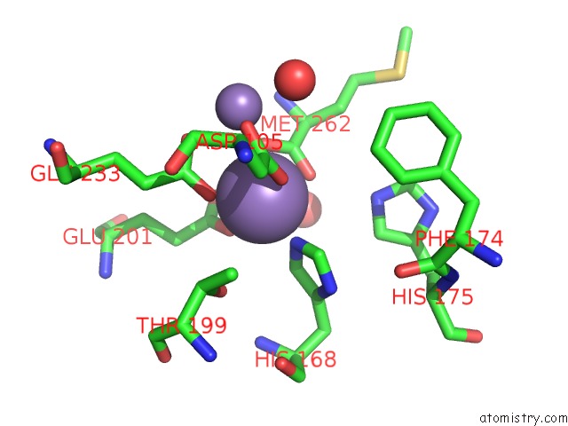
Manganese binding site 1 out of 4 in 3mx6
Go back to
Manganese binding site 1 out
of 4 in the Crystal Structure of Methionine Aminopeptidase From Rickettsia Prowazekii Bound to Methionine
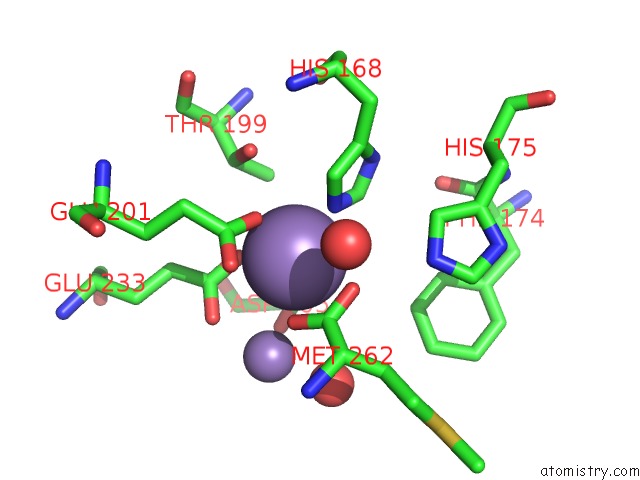
Mono view
Stereo pair view
Mono view
Stereo pair view
A full contact list of Manganese with other atoms in the Mn binding
site number 1 of Crystal Structure of Methionine Aminopeptidase From Rickettsia Prowazekii Bound to Methionine within 5.0Å range:
|
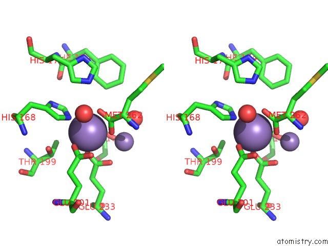
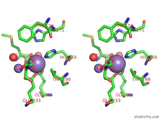
Manganese binding site 2 out of 4 in 3mx6
Go back to
Manganese binding site 2 out
of 4 in the Crystal Structure of Methionine Aminopeptidase From Rickettsia Prowazekii Bound to Methionine
Mono view
Stereo pair view
Mono view
Stereo pair view
A full contact list of Manganese with other atoms in the Mn binding
site number 2 of Crystal Structure of Methionine Aminopeptidase From Rickettsia Prowazekii Bound to Methionine within 5.0Å range:
|
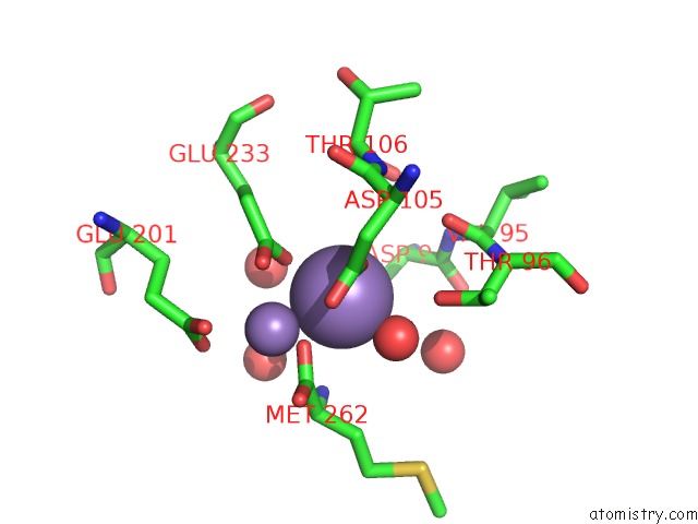
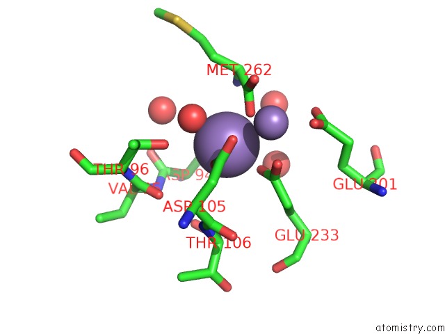
Manganese binding site 3 out of 4 in 3mx6
Go back to
Manganese binding site 3 out
of 4 in the Crystal Structure of Methionine Aminopeptidase From Rickettsia Prowazekii Bound to Methionine
Mono view
Stereo pair view
Mono view
Stereo pair view
A full contact list of Manganese with other atoms in the Mn binding
site number 3 of Crystal Structure of Methionine Aminopeptidase From Rickettsia Prowazekii Bound to Methionine within 5.0Å range:
|
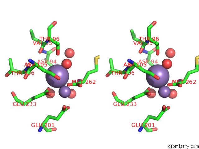
Manganese binding site 4 out of 4 in 3mx6
Go back to
Manganese binding site 4 out
of 4 in the Crystal Structure of Methionine Aminopeptidase From Rickettsia Prowazekii Bound to Methionine
Mono view
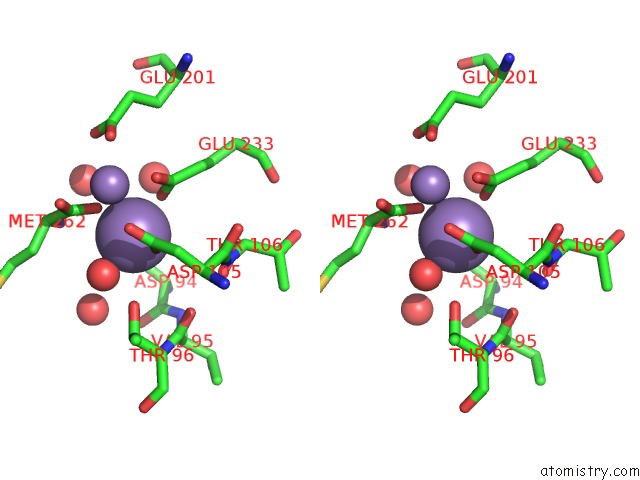
Stereo pair view
Mono view
Stereo pair view
A full contact list of Manganese with other atoms in the Mn binding
site number 4 of Crystal Structure of Methionine Aminopeptidase From Rickettsia Prowazekii Bound to Methionine within 5.0Å range:
|
Reference:
T.R.Helgren,
C.Chen,
P.Wangtrakuldee,
T.E.Edwards,
B.L.Staker,
J.Abendroth,
B.Sankaran,
N.A.Housley,
P.J.Myler,
J.P.Audia,
J.R.Horn,
T.J.Hagen.
Rickettsia Prowazekii Methionine Aminopeptidase As A Promising Target For the Development of Antibacterial Agents. Bioorg.Med.Chem. V. 25 813 2017.
ISSN: ISSN 0968-0896
PubMed: 28089350
DOI: 10.1016/J.BMC.2016.11.013
Page generated: Sat Oct 5 17:12:24 2024
ISSN: ISSN 0968-0896
PubMed: 28089350
DOI: 10.1016/J.BMC.2016.11.013
Last articles
Fe in 2YXOFe in 2YRS
Fe in 2YXC
Fe in 2YNM
Fe in 2YVJ
Fe in 2YP1
Fe in 2YU2
Fe in 2YU1
Fe in 2YQB
Fe in 2YOO