Manganese »
PDB 3m0m-3n37 »
3mwt »
Manganese in PDB 3mwt: Crystal Structure of Lassa Fever Virus Nucleoprotein in Complex with MN2+
Protein crystallography data
The structure of Crystal Structure of Lassa Fever Virus Nucleoprotein in Complex with MN2+, PDB code: 3mwt
was solved by
X.Qi,
S.Lan,
W.Wang,
L.M.Schelde,
H.Dong,
G.Wallat,
Y.Liang,
H.Ly,
C.Dong,
Scottish Structural Proteomics Facility (Sspf),
with X-Ray Crystallography technique. A brief refinement statistics is given in the table below:
| Resolution Low / High (Å) | 153.18 / 1.98 |
| Space group | P 3 |
| Cell size a, b, c (Å), α, β, γ (°) | 176.880, 176.880, 56.469, 90.00, 90.00, 120.00 |
| R / Rfree (%) | 18.1 / 21.8 |
Other elements in 3mwt:
The structure of Crystal Structure of Lassa Fever Virus Nucleoprotein in Complex with MN2+ also contains other interesting chemical elements:
| Zinc | (Zn) | 3 atoms |
Manganese Binding Sites:
The binding sites of Manganese atom in the Crystal Structure of Lassa Fever Virus Nucleoprotein in Complex with MN2+
(pdb code 3mwt). This binding sites where shown within
5.0 Angstroms radius around Manganese atom.
In total 3 binding sites of Manganese where determined in the Crystal Structure of Lassa Fever Virus Nucleoprotein in Complex with MN2+, PDB code: 3mwt:
Jump to Manganese binding site number: 1; 2; 3;
In total 3 binding sites of Manganese where determined in the Crystal Structure of Lassa Fever Virus Nucleoprotein in Complex with MN2+, PDB code: 3mwt:
Jump to Manganese binding site number: 1; 2; 3;
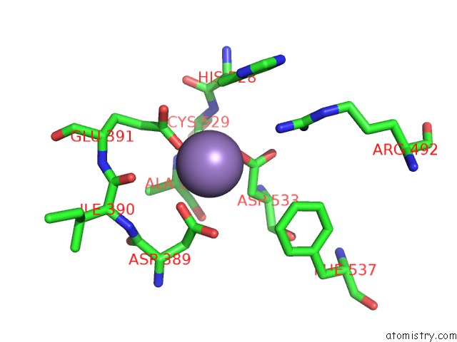
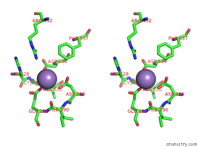
Manganese binding site 1 out of 3 in 3mwt
Go back to
Manganese binding site 1 out
of 3 in the Crystal Structure of Lassa Fever Virus Nucleoprotein in Complex with MN2+
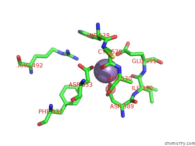
Mono view
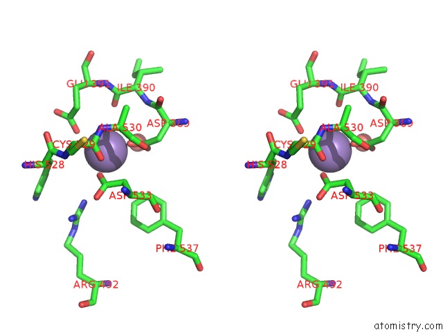
Stereo pair view
Mono view
Stereo pair view
A full contact list of Manganese with other atoms in the Mn binding
site number 1 of Crystal Structure of Lassa Fever Virus Nucleoprotein in Complex with MN2+ within 5.0Å range:
|
Manganese binding site 2 out of 3 in 3mwt
Go back to
Manganese binding site 2 out
of 3 in the Crystal Structure of Lassa Fever Virus Nucleoprotein in Complex with MN2+
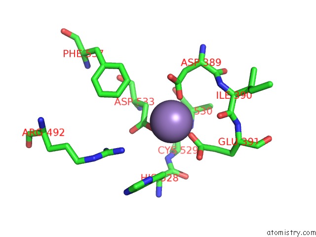
Mono view
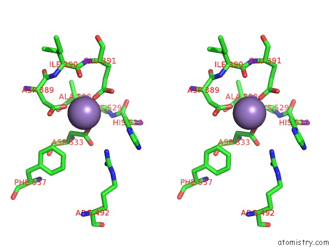
Stereo pair view
Mono view
Stereo pair view
A full contact list of Manganese with other atoms in the Mn binding
site number 2 of Crystal Structure of Lassa Fever Virus Nucleoprotein in Complex with MN2+ within 5.0Å range:
|
Manganese binding site 3 out of 3 in 3mwt
Go back to
Manganese binding site 3 out
of 3 in the Crystal Structure of Lassa Fever Virus Nucleoprotein in Complex with MN2+
Mono view
Stereo pair view
Mono view
Stereo pair view
A full contact list of Manganese with other atoms in the Mn binding
site number 3 of Crystal Structure of Lassa Fever Virus Nucleoprotein in Complex with MN2+ within 5.0Å range:
|
Reference:
X.Qi,
S.Lan,
W.Wang,
L.M.Schelde,
H.Dong,
G.D.Wallat,
H.Ly,
Y.Liang,
C.Dong.
Cap Binding and Immune Evasion Revealed By Lassa Nucleoprotein Structure. Nature V. 468 779 2010.
ISSN: ISSN 0028-0836
PubMed: 21085117
DOI: 10.1038/NATURE09605
Page generated: Sat Oct 5 17:12:21 2024
ISSN: ISSN 0028-0836
PubMed: 21085117
DOI: 10.1038/NATURE09605
Last articles
Zn in 9J0NZn in 9J0O
Zn in 9J0P
Zn in 9FJX
Zn in 9EKB
Zn in 9C0F
Zn in 9CAH
Zn in 9CH0
Zn in 9CH3
Zn in 9CH1