Manganese »
PDB 3m0m-3n37 »
3mof »
Manganese in PDB 3mof: The Structure of Rat Cytosolic Pepck Mutant A467G in Complex with Oxalate and Gtp
Enzymatic activity of The Structure of Rat Cytosolic Pepck Mutant A467G in Complex with Oxalate and Gtp
All present enzymatic activity of The Structure of Rat Cytosolic Pepck Mutant A467G in Complex with Oxalate and Gtp:
4.1.1.32;
4.1.1.32;
Protein crystallography data
The structure of The Structure of Rat Cytosolic Pepck Mutant A467G in Complex with Oxalate and Gtp, PDB code: 3mof
was solved by
T.A.Johnson,
T.Holyoak,
with X-Ray Crystallography technique. A brief refinement statistics is given in the table below:
| Resolution Low / High (Å) | 33.24 / 1.75 |
| Space group | P 1 21 1 |
| Cell size a, b, c (Å), α, β, γ (°) | 61.983, 119.601, 87.068, 90.00, 106.87, 90.00 |
| R / Rfree (%) | 17.9 / 22.8 |
Other elements in 3mof:
The structure of The Structure of Rat Cytosolic Pepck Mutant A467G in Complex with Oxalate and Gtp also contains other interesting chemical elements:
| Sodium | (Na) | 2 atoms |
Manganese Binding Sites:
The binding sites of Manganese atom in the The Structure of Rat Cytosolic Pepck Mutant A467G in Complex with Oxalate and Gtp
(pdb code 3mof). This binding sites where shown within
5.0 Angstroms radius around Manganese atom.
In total 5 binding sites of Manganese where determined in the The Structure of Rat Cytosolic Pepck Mutant A467G in Complex with Oxalate and Gtp, PDB code: 3mof:
Jump to Manganese binding site number: 1; 2; 3; 4; 5;
In total 5 binding sites of Manganese where determined in the The Structure of Rat Cytosolic Pepck Mutant A467G in Complex with Oxalate and Gtp, PDB code: 3mof:
Jump to Manganese binding site number: 1; 2; 3; 4; 5;
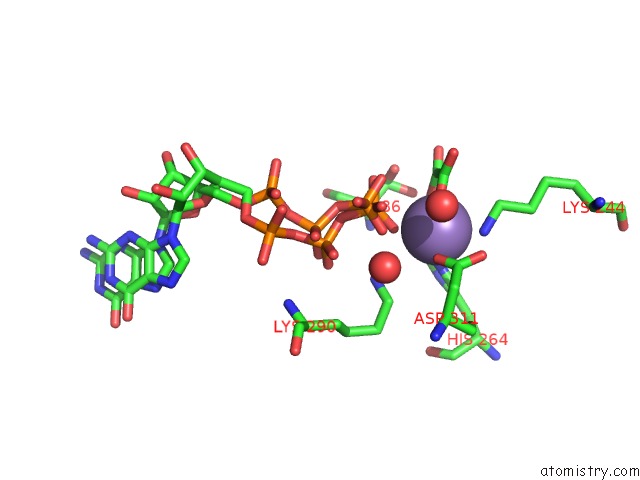
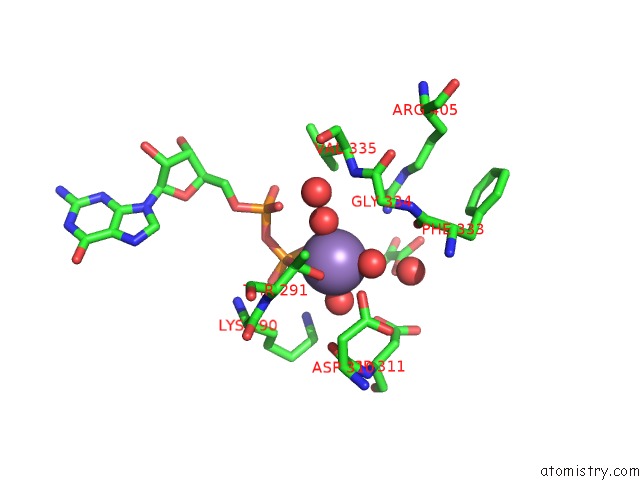
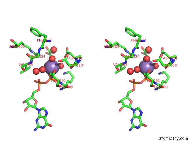
Manganese binding site 1 out of 5 in 3mof
Go back to
Manganese binding site 1 out
of 5 in the The Structure of Rat Cytosolic Pepck Mutant A467G in Complex with Oxalate and Gtp
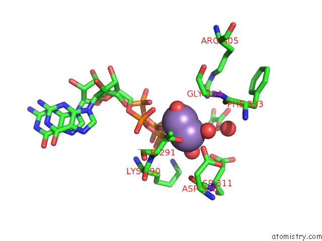
Mono view
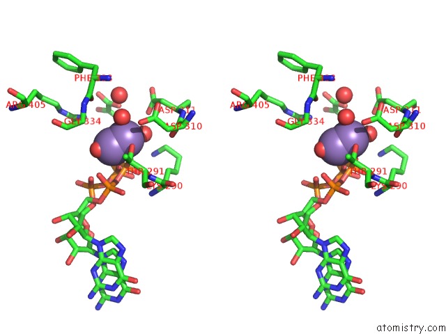
Stereo pair view
Mono view
Stereo pair view
A full contact list of Manganese with other atoms in the Mn binding
site number 1 of The Structure of Rat Cytosolic Pepck Mutant A467G in Complex with Oxalate and Gtp within 5.0Å range:
|
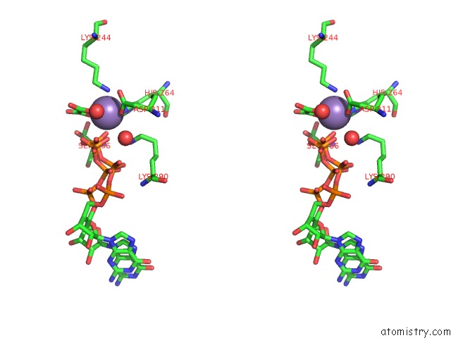
Manganese binding site 2 out of 5 in 3mof
Go back to
Manganese binding site 2 out
of 5 in the The Structure of Rat Cytosolic Pepck Mutant A467G in Complex with Oxalate and Gtp
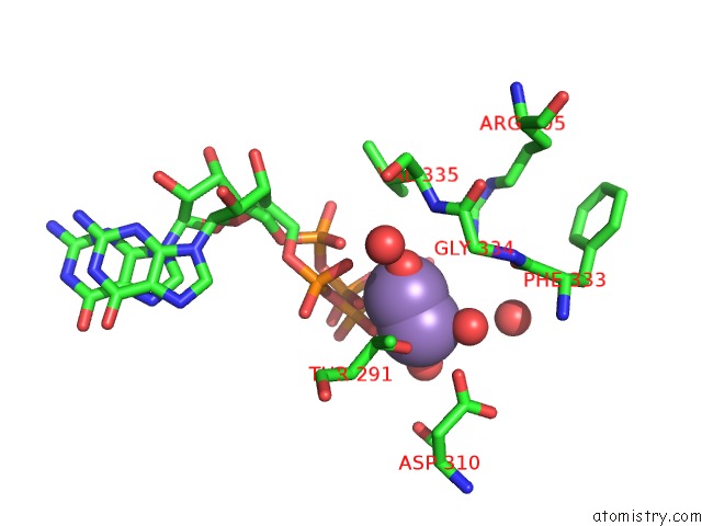
Mono view
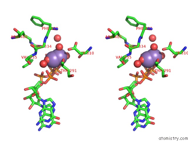
Stereo pair view
Mono view
Stereo pair view
A full contact list of Manganese with other atoms in the Mn binding
site number 2 of The Structure of Rat Cytosolic Pepck Mutant A467G in Complex with Oxalate and Gtp within 5.0Å range:
|
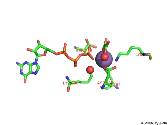
Manganese binding site 3 out of 5 in 3mof
Go back to
Manganese binding site 3 out
of 5 in the The Structure of Rat Cytosolic Pepck Mutant A467G in Complex with Oxalate and Gtp
Mono view
Stereo pair view
Mono view
Stereo pair view
A full contact list of Manganese with other atoms in the Mn binding
site number 3 of The Structure of Rat Cytosolic Pepck Mutant A467G in Complex with Oxalate and Gtp within 5.0Å range:
|
Manganese binding site 4 out of 5 in 3mof
Go back to
Manganese binding site 4 out
of 5 in the The Structure of Rat Cytosolic Pepck Mutant A467G in Complex with Oxalate and Gtp
Mono view
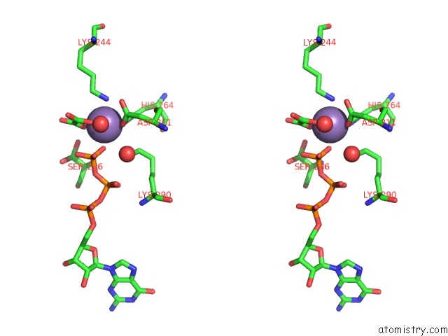
Stereo pair view
Mono view
Stereo pair view
A full contact list of Manganese with other atoms in the Mn binding
site number 4 of The Structure of Rat Cytosolic Pepck Mutant A467G in Complex with Oxalate and Gtp within 5.0Å range:
|
Manganese binding site 5 out of 5 in 3mof
Go back to
Manganese binding site 5 out
of 5 in the The Structure of Rat Cytosolic Pepck Mutant A467G in Complex with Oxalate and Gtp
Mono view
Stereo pair view
Mono view
Stereo pair view
A full contact list of Manganese with other atoms in the Mn binding
site number 5 of The Structure of Rat Cytosolic Pepck Mutant A467G in Complex with Oxalate and Gtp within 5.0Å range:
|
Reference:
T.A.Johnson,
T.Holyoak.
Increasing the Conformational Entropy of the Omega-Loop Lid Domain in Phosphoenolpyruvate Carboxykinase Impairs Catalysis and Decreases Catalytic Fidelity . Biochemistry V. 49 5176 2010.
ISSN: ISSN 0006-2960
PubMed: 20476774
DOI: 10.1021/BI100399E
Page generated: Sat Oct 5 17:09:57 2024
ISSN: ISSN 0006-2960
PubMed: 20476774
DOI: 10.1021/BI100399E
Last articles
As in 4BKSAs in 3WGH
As in 3ZPG
As in 3W88
As in 3W87
As in 3WE3
As in 3WC5
As in 3WAB
As in 3UJP
As in 3W86