Manganese »
PDB 3kky-3m0l »
3m0j »
Manganese in PDB 3m0j: Structure of Oxaloacetate Acetylhydrolase in Complex with the Inhibitor 3,3-Difluorooxalacetate
Enzymatic activity of Structure of Oxaloacetate Acetylhydrolase in Complex with the Inhibitor 3,3-Difluorooxalacetate
All present enzymatic activity of Structure of Oxaloacetate Acetylhydrolase in Complex with the Inhibitor 3,3-Difluorooxalacetate:
3.7.1.1;
3.7.1.1;
Protein crystallography data
The structure of Structure of Oxaloacetate Acetylhydrolase in Complex with the Inhibitor 3,3-Difluorooxalacetate, PDB code: 3m0j
was solved by
O.Herzberg,
C.Chen,
with X-Ray Crystallography technique. A brief refinement statistics is given in the table below:
| Resolution Low / High (Å) | 19.92 / 1.55 |
| Space group | P 42 21 2 |
| Cell size a, b, c (Å), α, β, γ (°) | 82.144, 82.144, 72.660, 90.00, 90.00, 90.00 |
| R / Rfree (%) | 17.5 / 20.1 |
Other elements in 3m0j:
The structure of Structure of Oxaloacetate Acetylhydrolase in Complex with the Inhibitor 3,3-Difluorooxalacetate also contains other interesting chemical elements:
| Fluorine | (F) | 2 atoms |
| Calcium | (Ca) | 1 atom |
Manganese Binding Sites:
The binding sites of Manganese atom in the Structure of Oxaloacetate Acetylhydrolase in Complex with the Inhibitor 3,3-Difluorooxalacetate
(pdb code 3m0j). This binding sites where shown within
5.0 Angstroms radius around Manganese atom.
In total only one binding site of Manganese was determined in the Structure of Oxaloacetate Acetylhydrolase in Complex with the Inhibitor 3,3-Difluorooxalacetate, PDB code: 3m0j:
In total only one binding site of Manganese was determined in the Structure of Oxaloacetate Acetylhydrolase in Complex with the Inhibitor 3,3-Difluorooxalacetate, PDB code: 3m0j:
Manganese binding site 1 out of 1 in 3m0j
Go back to
Manganese binding site 1 out
of 1 in the Structure of Oxaloacetate Acetylhydrolase in Complex with the Inhibitor 3,3-Difluorooxalacetate
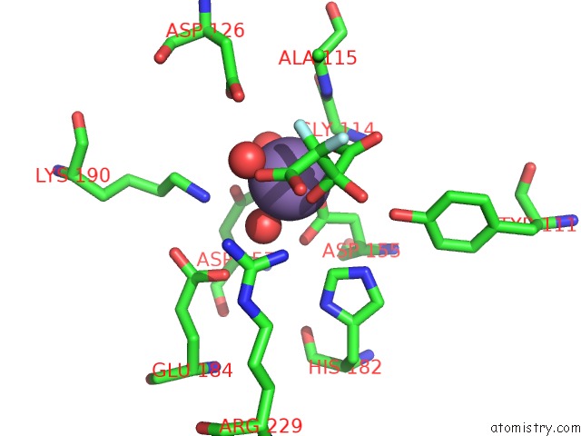
Mono view
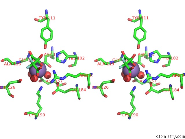
Stereo pair view
Mono view
Stereo pair view
A full contact list of Manganese with other atoms in the Mn binding
site number 1 of Structure of Oxaloacetate Acetylhydrolase in Complex with the Inhibitor 3,3-Difluorooxalacetate within 5.0Å range:
|
Reference:
C.Chen,
Q.Sun,
B.Narayanan,
D.L.Nuss,
O.Herzberg.
Structure of Oxalacetate Acetylhydrolase, A Virulence Factor of the Chestnut Blight Fungus. J.Biol.Chem. V. 285 26685 2010.
ISSN: ISSN 0021-9258
PubMed: 20558740
DOI: 10.1074/JBC.M110.117804
Page generated: Sat Oct 5 16:55:22 2024
ISSN: ISSN 0021-9258
PubMed: 20558740
DOI: 10.1074/JBC.M110.117804
Last articles
Zn in 9J0NZn in 9J0O
Zn in 9J0P
Zn in 9FJX
Zn in 9EKB
Zn in 9C0F
Zn in 9CAH
Zn in 9CH0
Zn in 9CH3
Zn in 9CH1