Manganese »
PDB 3kky-3m0l »
3lz0 »
Manganese in PDB 3lz0: Crystal Structure of Nucleosome Core Particle Composed of the Widom 601 Dna Sequence (Orientation 1)
Protein crystallography data
The structure of Crystal Structure of Nucleosome Core Particle Composed of the Widom 601 Dna Sequence (Orientation 1), PDB code: 3lz0
was solved by
D.Vasudevan,
E.Y.D.Chua,
C.A.Davey,
with X-Ray Crystallography technique. A brief refinement statistics is given in the table below:
| Resolution Low / High (Å) | 93.04 / 2.50 |
| Space group | P 21 21 21 |
| Cell size a, b, c (Å), α, β, γ (°) | 107.370, 109.660, 175.750, 90.00, 90.00, 90.00 |
| R / Rfree (%) | 26.8 / 31.8 |
Other elements in 3lz0:
The structure of Crystal Structure of Nucleosome Core Particle Composed of the Widom 601 Dna Sequence (Orientation 1) also contains other interesting chemical elements:
| Chlorine | (Cl) | 2 atoms |
Manganese Binding Sites:
The binding sites of Manganese atom in the Crystal Structure of Nucleosome Core Particle Composed of the Widom 601 Dna Sequence (Orientation 1)
(pdb code 3lz0). This binding sites where shown within
5.0 Angstroms radius around Manganese atom.
In total 8 binding sites of Manganese where determined in the Crystal Structure of Nucleosome Core Particle Composed of the Widom 601 Dna Sequence (Orientation 1), PDB code: 3lz0:
Jump to Manganese binding site number: 1; 2; 3; 4; 5; 6; 7; 8;
In total 8 binding sites of Manganese where determined in the Crystal Structure of Nucleosome Core Particle Composed of the Widom 601 Dna Sequence (Orientation 1), PDB code: 3lz0:
Jump to Manganese binding site number: 1; 2; 3; 4; 5; 6; 7; 8;
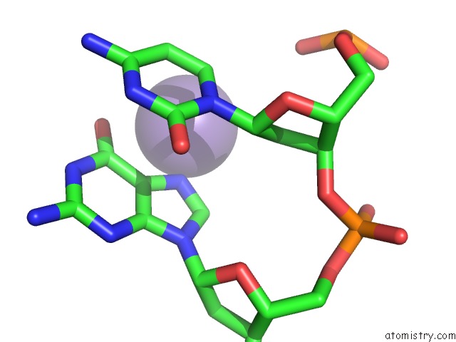
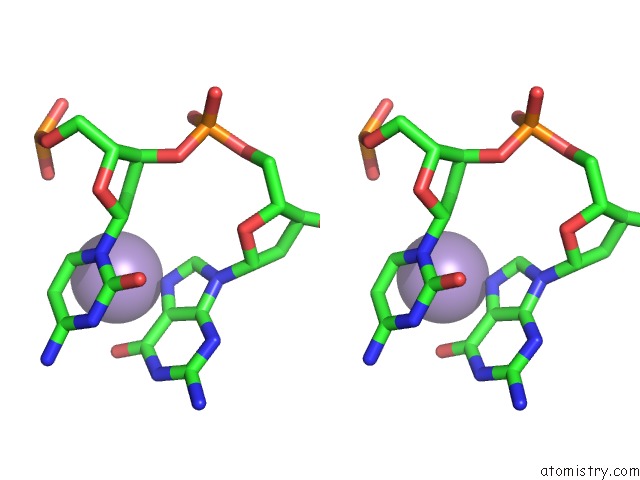
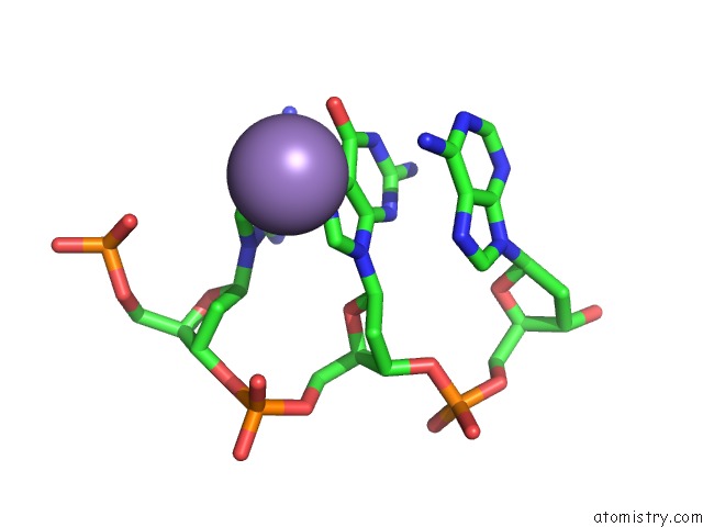
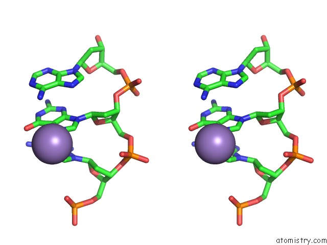
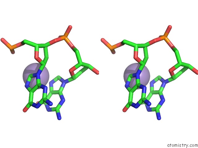
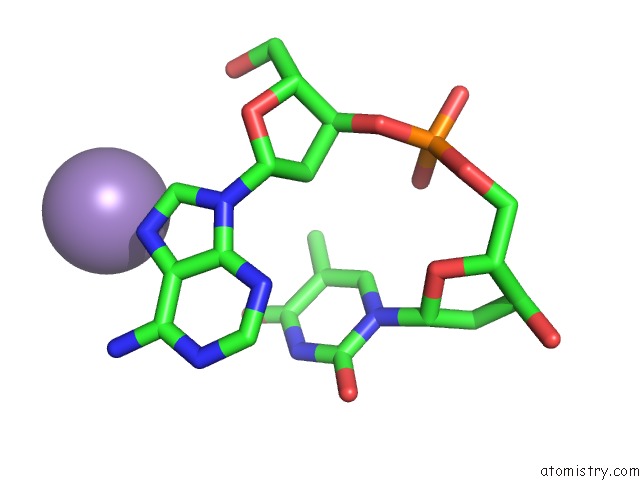
Manganese binding site 1 out of 8 in 3lz0
Go back to
Manganese binding site 1 out
of 8 in the Crystal Structure of Nucleosome Core Particle Composed of the Widom 601 Dna Sequence (Orientation 1)
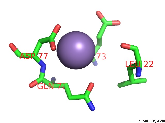
Mono view
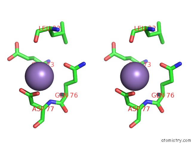
Stereo pair view
Mono view
Stereo pair view
A full contact list of Manganese with other atoms in the Mn binding
site number 1 of Crystal Structure of Nucleosome Core Particle Composed of the Widom 601 Dna Sequence (Orientation 1) within 5.0Å range:
|
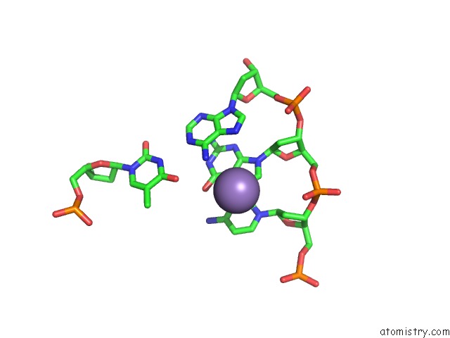
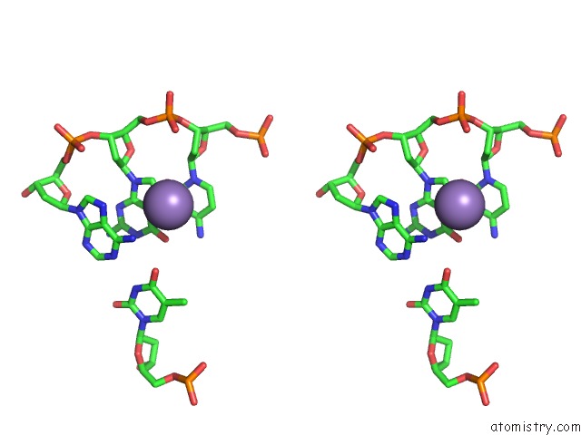
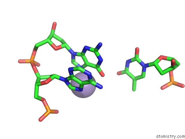
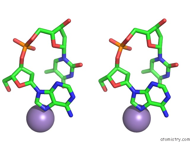
Manganese binding site 2 out of 8 in 3lz0
Go back to
Manganese binding site 2 out
of 8 in the Crystal Structure of Nucleosome Core Particle Composed of the Widom 601 Dna Sequence (Orientation 1)
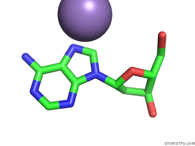
Mono view
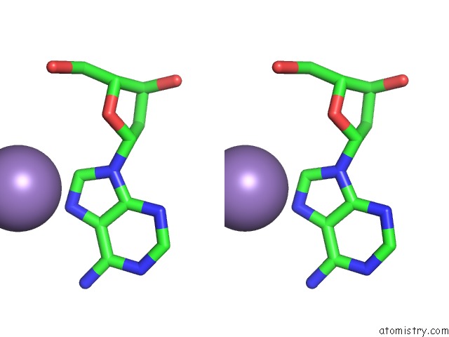
Stereo pair view
Mono view
Stereo pair view
A full contact list of Manganese with other atoms in the Mn binding
site number 2 of Crystal Structure of Nucleosome Core Particle Composed of the Widom 601 Dna Sequence (Orientation 1) within 5.0Å range:
|
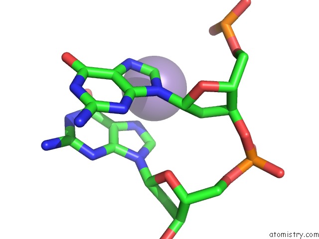
Manganese binding site 3 out of 8 in 3lz0
Go back to
Manganese binding site 3 out
of 8 in the Crystal Structure of Nucleosome Core Particle Composed of the Widom 601 Dna Sequence (Orientation 1)
Mono view
Stereo pair view
Mono view
Stereo pair view
A full contact list of Manganese with other atoms in the Mn binding
site number 3 of Crystal Structure of Nucleosome Core Particle Composed of the Widom 601 Dna Sequence (Orientation 1) within 5.0Å range:
|
Manganese binding site 4 out of 8 in 3lz0
Go back to
Manganese binding site 4 out
of 8 in the Crystal Structure of Nucleosome Core Particle Composed of the Widom 601 Dna Sequence (Orientation 1)
Mono view
Stereo pair view
Mono view
Stereo pair view
A full contact list of Manganese with other atoms in the Mn binding
site number 4 of Crystal Structure of Nucleosome Core Particle Composed of the Widom 601 Dna Sequence (Orientation 1) within 5.0Å range:
|
Manganese binding site 5 out of 8 in 3lz0
Go back to
Manganese binding site 5 out
of 8 in the Crystal Structure of Nucleosome Core Particle Composed of the Widom 601 Dna Sequence (Orientation 1)
Mono view
Stereo pair view
Mono view
Stereo pair view
A full contact list of Manganese with other atoms in the Mn binding
site number 5 of Crystal Structure of Nucleosome Core Particle Composed of the Widom 601 Dna Sequence (Orientation 1) within 5.0Å range:
|
Manganese binding site 6 out of 8 in 3lz0
Go back to
Manganese binding site 6 out
of 8 in the Crystal Structure of Nucleosome Core Particle Composed of the Widom 601 Dna Sequence (Orientation 1)
Mono view

Stereo pair view
Mono view
Stereo pair view
A full contact list of Manganese with other atoms in the Mn binding
site number 6 of Crystal Structure of Nucleosome Core Particle Composed of the Widom 601 Dna Sequence (Orientation 1) within 5.0Å range:
|
Manganese binding site 7 out of 8 in 3lz0
Go back to
Manganese binding site 7 out
of 8 in the Crystal Structure of Nucleosome Core Particle Composed of the Widom 601 Dna Sequence (Orientation 1)
Mono view
Stereo pair view
Mono view
Stereo pair view
A full contact list of Manganese with other atoms in the Mn binding
site number 7 of Crystal Structure of Nucleosome Core Particle Composed of the Widom 601 Dna Sequence (Orientation 1) within 5.0Å range:
|
Manganese binding site 8 out of 8 in 3lz0
Go back to
Manganese binding site 8 out
of 8 in the Crystal Structure of Nucleosome Core Particle Composed of the Widom 601 Dna Sequence (Orientation 1)
Mono view
Stereo pair view
Mono view
Stereo pair view
A full contact list of Manganese with other atoms in the Mn binding
site number 8 of Crystal Structure of Nucleosome Core Particle Composed of the Widom 601 Dna Sequence (Orientation 1) within 5.0Å range:
|
Reference:
D.Vasudevan,
E.Y.Chua,
C.A.Davey.
Crystal Structures of Nucleosome Core Particles Containing the '601' Strong Positioning Sequence J.Mol.Biol. V. 403 1 2010.
ISSN: ISSN 0022-2836
PubMed: 20800598
DOI: 10.1016/J.JMB.2010.08.039
Page generated: Sat Oct 5 16:53:24 2024
ISSN: ISSN 0022-2836
PubMed: 20800598
DOI: 10.1016/J.JMB.2010.08.039
Last articles
Zn in 9J0NZn in 9J0O
Zn in 9J0P
Zn in 9FJX
Zn in 9EKB
Zn in 9C0F
Zn in 9CAH
Zn in 9CH0
Zn in 9CH3
Zn in 9CH1