Manganese »
PDB 3kky-3m0l »
3lw6 »
Manganese in PDB 3lw6: Crystal Structure of Drosophila BETA1,4-Galactosyltransferase-7
Enzymatic activity of Crystal Structure of Drosophila BETA1,4-Galactosyltransferase-7
All present enzymatic activity of Crystal Structure of Drosophila BETA1,4-Galactosyltransferase-7:
2.4.1.133;
2.4.1.133;
Protein crystallography data
The structure of Crystal Structure of Drosophila BETA1,4-Galactosyltransferase-7, PDB code: 3lw6
was solved by
B.Ramakrishnan,
P.K.Qasba,
with X-Ray Crystallography technique. A brief refinement statistics is given in the table below:
| Resolution Low / High (Å) | 36.60 / 1.81 |
| Space group | P 43 21 2 |
| Cell size a, b, c (Å), α, β, γ (°) | 81.835, 81.835, 133.656, 90.00, 90.00, 90.00 |
| R / Rfree (%) | 18.9 / 22.1 |
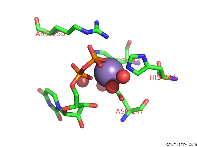
Manganese Binding Sites:
The binding sites of Manganese atom in the Crystal Structure of Drosophila BETA1,4-Galactosyltransferase-7
(pdb code 3lw6). This binding sites where shown within
5.0 Angstroms radius around Manganese atom.
In total only one binding site of Manganese was determined in the Crystal Structure of Drosophila BETA1,4-Galactosyltransferase-7, PDB code: 3lw6:
In total only one binding site of Manganese was determined in the Crystal Structure of Drosophila BETA1,4-Galactosyltransferase-7, PDB code: 3lw6:
Manganese binding site 1 out of 1 in 3lw6
Go back to
Manganese binding site 1 out
of 1 in the Crystal Structure of Drosophila BETA1,4-Galactosyltransferase-7
Mono view

Stereo pair view
Mono view
Stereo pair view
A full contact list of Manganese with other atoms in the Mn binding
site number 1 of Crystal Structure of Drosophila BETA1,4-Galactosyltransferase-7 within 5.0Å range:
|
Reference:
B.Ramakrishnan,
P.K.Qasba.
Crystal Structure of the Catalytic Domain of Drosophila BETA1,4-Galactosyltransferase-7. J.Biol.Chem. V. 285 15619 2010.
ISSN: ISSN 0021-9258
PubMed: 20236943
DOI: 10.1074/JBC.M109.099564
Page generated: Sat Aug 16 12:12:28 2025
ISSN: ISSN 0021-9258
PubMed: 20236943
DOI: 10.1074/JBC.M109.099564
Last articles
Mn in 4LPFMn in 4LOM
Mn in 4L6D
Mn in 4LNC
Mn in 4LN7
Mn in 4LFI
Mn in 4LIL
Mn in 4LAC
Mn in 4L8Q
Mn in 4L78