Manganese »
PDB 3kky-3m0l »
3lp4 »
Manganese in PDB 3lp4: Crystal Structure of Human Arginase I in Complex with L-Lysine, 1.90A Resolution.
Enzymatic activity of Crystal Structure of Human Arginase I in Complex with L-Lysine, 1.90A Resolution.
All present enzymatic activity of Crystal Structure of Human Arginase I in Complex with L-Lysine, 1.90A Resolution.:
3.5.3.1;
3.5.3.1;
Protein crystallography data
The structure of Crystal Structure of Human Arginase I in Complex with L-Lysine, 1.90A Resolution., PDB code: 3lp4
was solved by
L.Di Costanzo,
D.W.Christianson,
with X-Ray Crystallography technique. A brief refinement statistics is given in the table below:
| Resolution Low / High (Å) | 34.20 / 1.90 |
| Space group | P 3 |
| Cell size a, b, c (Å), α, β, γ (°) | 90.729, 90.729, 69.509, 90.00, 90.00, 120.00 |
| R / Rfree (%) | 15.5 / 20.4 |
Manganese Binding Sites:
The binding sites of Manganese atom in the Crystal Structure of Human Arginase I in Complex with L-Lysine, 1.90A Resolution.
(pdb code 3lp4). This binding sites where shown within
5.0 Angstroms radius around Manganese atom.
In total 4 binding sites of Manganese where determined in the Crystal Structure of Human Arginase I in Complex with L-Lysine, 1.90A Resolution., PDB code: 3lp4:
Jump to Manganese binding site number: 1; 2; 3; 4;
In total 4 binding sites of Manganese where determined in the Crystal Structure of Human Arginase I in Complex with L-Lysine, 1.90A Resolution., PDB code: 3lp4:
Jump to Manganese binding site number: 1; 2; 3; 4;
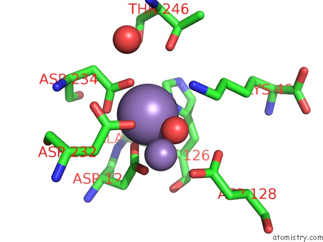
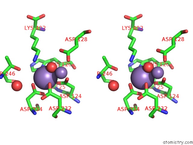
Manganese binding site 1 out of 4 in 3lp4
Go back to
Manganese binding site 1 out
of 4 in the Crystal Structure of Human Arginase I in Complex with L-Lysine, 1.90A Resolution.
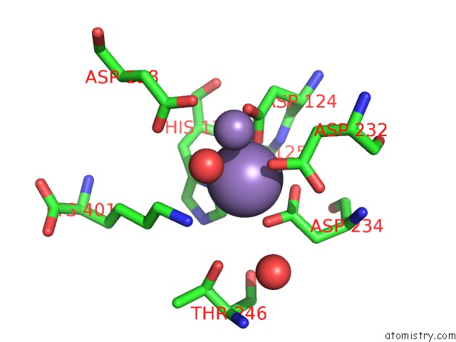
Mono view
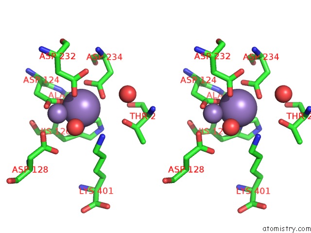
Stereo pair view
Mono view
Stereo pair view
A full contact list of Manganese with other atoms in the Mn binding
site number 1 of Crystal Structure of Human Arginase I in Complex with L-Lysine, 1.90A Resolution. within 5.0Å range:
|
Manganese binding site 2 out of 4 in 3lp4
Go back to
Manganese binding site 2 out
of 4 in the Crystal Structure of Human Arginase I in Complex with L-Lysine, 1.90A Resolution.
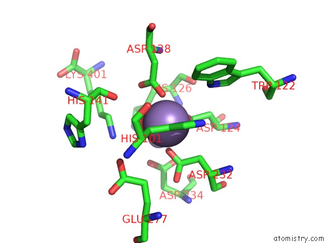
Mono view
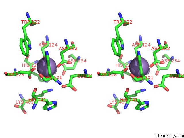
Stereo pair view
Mono view
Stereo pair view
A full contact list of Manganese with other atoms in the Mn binding
site number 2 of Crystal Structure of Human Arginase I in Complex with L-Lysine, 1.90A Resolution. within 5.0Å range:
|
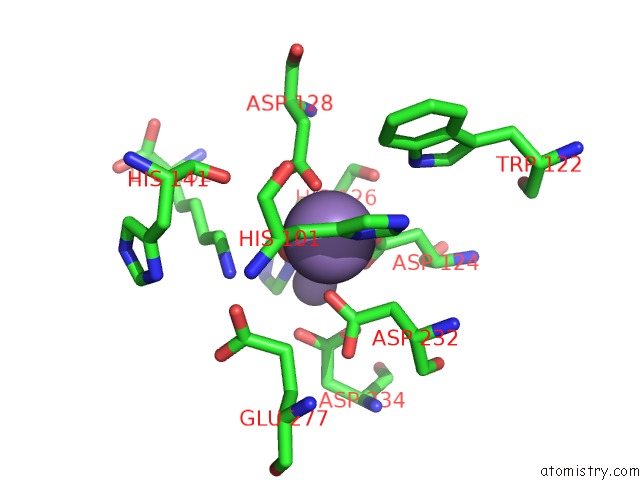
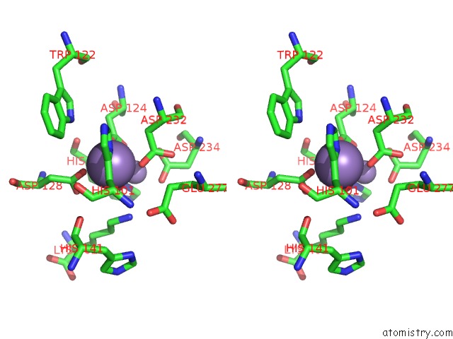
Manganese binding site 3 out of 4 in 3lp4
Go back to
Manganese binding site 3 out
of 4 in the Crystal Structure of Human Arginase I in Complex with L-Lysine, 1.90A Resolution.
Mono view
Stereo pair view
Mono view
Stereo pair view
A full contact list of Manganese with other atoms in the Mn binding
site number 3 of Crystal Structure of Human Arginase I in Complex with L-Lysine, 1.90A Resolution. within 5.0Å range:
|
Manganese binding site 4 out of 4 in 3lp4
Go back to
Manganese binding site 4 out
of 4 in the Crystal Structure of Human Arginase I in Complex with L-Lysine, 1.90A Resolution.
Mono view
Stereo pair view
Mono view
Stereo pair view
A full contact list of Manganese with other atoms in the Mn binding
site number 4 of Crystal Structure of Human Arginase I in Complex with L-Lysine, 1.90A Resolution. within 5.0Å range:
|
Reference:
L.Di Costanzo,
M.Ilies,
K.J.Thorn,
D.W.Christianson.
Inhibition of Human Arginase I By Substrate and Product Analogues. Arch.Biochem.Biophys. V. 496 101 2010.
ISSN: ISSN 0003-9861
PubMed: 20153713
DOI: 10.1016/J.ABB.2010.02.004
Page generated: Sat Oct 5 16:52:14 2024
ISSN: ISSN 0003-9861
PubMed: 20153713
DOI: 10.1016/J.ABB.2010.02.004
Last articles
Zn in 9J0NZn in 9J0O
Zn in 9J0P
Zn in 9FJX
Zn in 9EKB
Zn in 9C0F
Zn in 9CAH
Zn in 9CH0
Zn in 9CH3
Zn in 9CH1