Manganese »
PDB 3kky-3m0l »
3loj »
Manganese in PDB 3loj: Structure of Mycobacterium Tuberculosis Dutpase H145A Mutant
Enzymatic activity of Structure of Mycobacterium Tuberculosis Dutpase H145A Mutant
All present enzymatic activity of Structure of Mycobacterium Tuberculosis Dutpase H145A Mutant:
3.6.1.23;
3.6.1.23;
Protein crystallography data
The structure of Structure of Mycobacterium Tuberculosis Dutpase H145A Mutant, PDB code: 3loj
was solved by
I.Leveles,
V.Harmat,
I.Pecsi,
A.Lopata,
B.G.Vertessy,
J.Toth,
with X-Ray Crystallography technique. A brief refinement statistics is given in the table below:
| Resolution Low / High (Å) | 30.00 / 1.25 |
| Space group | P 63 |
| Cell size a, b, c (Å), α, β, γ (°) | 54.591, 54.591, 83.051, 90.00, 90.00, 120.00 |
| R / Rfree (%) | 12.8 / 17.7 |
Other elements in 3loj:
The structure of Structure of Mycobacterium Tuberculosis Dutpase H145A Mutant also contains other interesting chemical elements:
| Magnesium | (Mg) | 1 atom |
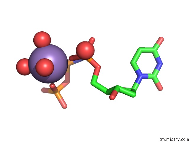
Manganese Binding Sites:
The binding sites of Manganese atom in the Structure of Mycobacterium Tuberculosis Dutpase H145A Mutant
(pdb code 3loj). This binding sites where shown within
5.0 Angstroms radius around Manganese atom.
In total only one binding site of Manganese was determined in the Structure of Mycobacterium Tuberculosis Dutpase H145A Mutant, PDB code: 3loj:
In total only one binding site of Manganese was determined in the Structure of Mycobacterium Tuberculosis Dutpase H145A Mutant, PDB code: 3loj:
Manganese binding site 1 out of 1 in 3loj
Go back to
Manganese binding site 1 out
of 1 in the Structure of Mycobacterium Tuberculosis Dutpase H145A Mutant
Mono view

Stereo pair view
Mono view
Stereo pair view
A full contact list of Manganese with other atoms in the Mn binding
site number 1 of Structure of Mycobacterium Tuberculosis Dutpase H145A Mutant within 5.0Å range:
|
Reference:
I.Pecsi,
I.Leveles,
V.Harmat,
B.G.Vertessy,
J.Toth.
Aromatic Stacking Between Nucleobase and Enzyme Promotes Phosphate Ester Hydrolysis in Dutpase Nucleic Acids Res. V. 38 7179 2010.
ISSN: ISSN 0305-1048
PubMed: 20601405
DOI: 10.1093/NAR/GKQ584
Page generated: Sat Oct 5 16:50:25 2024
ISSN: ISSN 0305-1048
PubMed: 20601405
DOI: 10.1093/NAR/GKQ584
Last articles
Zn in 9J0NZn in 9J0O
Zn in 9J0P
Zn in 9FJX
Zn in 9EKB
Zn in 9C0F
Zn in 9CAH
Zn in 9CH0
Zn in 9CH3
Zn in 9CH1