Manganese »
PDB 3hw6-3ki9 »
3ju9 »
Manganese in PDB 3ju9: Crystal Structure of A Lectin From Canavalia Brasiliensis Seed (Conbr) Complexed with Alpha-Aminobutyric Acid
Protein crystallography data
The structure of Crystal Structure of A Lectin From Canavalia Brasiliensis Seed (Conbr) Complexed with Alpha-Aminobutyric Acid, PDB code: 3ju9
was solved by
E.H.S.Bezerra,
B.A.M.Rocha,
C.S.Nagano,
G.A.Bezerra,
T.R.Moura,
M.J.B.Bezerra,
R.G.Benevides,
E.S.Marinho,
P.Delatorre,
B.S.Cavada,
with X-Ray Crystallography technique. A brief refinement statistics is given in the table below:
| Resolution Low / High (Å) | 36.51 / 2.10 |
| Space group | I 2 2 2 |
| Cell size a, b, c (Å), α, β, γ (°) | 68.323, 73.020, 99.542, 90.00, 90.00, 90.00 |
| R / Rfree (%) | 20.4 / 25.3 |
Other elements in 3ju9:
The structure of Crystal Structure of A Lectin From Canavalia Brasiliensis Seed (Conbr) Complexed with Alpha-Aminobutyric Acid also contains other interesting chemical elements:
| Chlorine | (Cl) | 2 atoms |
| Calcium | (Ca) | 1 atom |
Manganese Binding Sites:
The binding sites of Manganese atom in the Crystal Structure of A Lectin From Canavalia Brasiliensis Seed (Conbr) Complexed with Alpha-Aminobutyric Acid
(pdb code 3ju9). This binding sites where shown within
5.0 Angstroms radius around Manganese atom.
In total only one binding site of Manganese was determined in the Crystal Structure of A Lectin From Canavalia Brasiliensis Seed (Conbr) Complexed with Alpha-Aminobutyric Acid, PDB code: 3ju9:
In total only one binding site of Manganese was determined in the Crystal Structure of A Lectin From Canavalia Brasiliensis Seed (Conbr) Complexed with Alpha-Aminobutyric Acid, PDB code: 3ju9:
Manganese binding site 1 out of 1 in 3ju9
Go back to
Manganese binding site 1 out
of 1 in the Crystal Structure of A Lectin From Canavalia Brasiliensis Seed (Conbr) Complexed with Alpha-Aminobutyric Acid
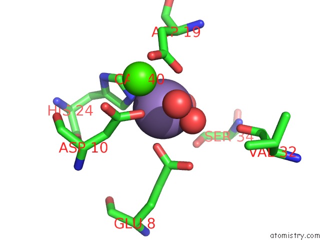
Mono view
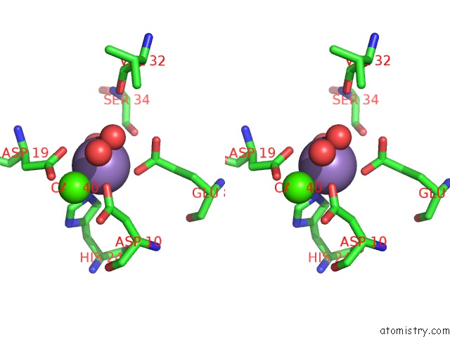
Stereo pair view
Mono view
Stereo pair view
A full contact list of Manganese with other atoms in the Mn binding
site number 1 of Crystal Structure of A Lectin From Canavalia Brasiliensis Seed (Conbr) Complexed with Alpha-Aminobutyric Acid within 5.0Å range:
|
Reference:
E.H.Bezerra,
B.A.Rocha,
C.S.Nagano,
G.A.Bezerra,
T.R.Moura,
M.J.Bezerra,
R.G.Benevides,
A.H.Sampaio,
A.M.Assreuy,
P.Delatorre,
B.S.Cavada.
Structural Analysis of Conbr Reveals Molecular Correlation Between the Carbohydrate Recognition Domain and Endothelial No Synthase Activation. Biochem.Biophys.Res.Commun. V. 408 566 2011.
ISSN: ISSN 0006-291X
PubMed: 21530490
DOI: 10.1016/J.BBRC.2011.04.061
Page generated: Sat Oct 5 16:42:14 2024
ISSN: ISSN 0006-291X
PubMed: 21530490
DOI: 10.1016/J.BBRC.2011.04.061
Last articles
Zn in 9MJ5Zn in 9HNW
Zn in 9G0L
Zn in 9FNE
Zn in 9DZN
Zn in 9E0I
Zn in 9D32
Zn in 9DAK
Zn in 8ZXC
Zn in 8ZUF