Manganese »
PDB 3hw6-3ki9 »
3ito »
Manganese in PDB 3ito: Crystal Structure of Pseudomonas Stutzeri L-Rhamnose Isomerase Mutant D327N in Complex with D-Psicose
Enzymatic activity of Crystal Structure of Pseudomonas Stutzeri L-Rhamnose Isomerase Mutant D327N in Complex with D-Psicose
All present enzymatic activity of Crystal Structure of Pseudomonas Stutzeri L-Rhamnose Isomerase Mutant D327N in Complex with D-Psicose:
5.3.1.14;
5.3.1.14;
Protein crystallography data
The structure of Crystal Structure of Pseudomonas Stutzeri L-Rhamnose Isomerase Mutant D327N in Complex with D-Psicose, PDB code: 3ito
was solved by
H.Yoshida,
M.Yamaji,
T.Ishii,
K.Izumori,
S.Kamitori,
with X-Ray Crystallography technique. A brief refinement statistics is given in the table below:
| Resolution Low / High (Å) | 42.15 / 1.90 |
| Space group | P 1 21 1 |
| Cell size a, b, c (Å), α, β, γ (°) | 74.762, 104.732, 115.035, 90.00, 108.24, 90.00 |
| R / Rfree (%) | 17.6 / 21.1 |
Manganese Binding Sites:
The binding sites of Manganese atom in the Crystal Structure of Pseudomonas Stutzeri L-Rhamnose Isomerase Mutant D327N in Complex with D-Psicose
(pdb code 3ito). This binding sites where shown within
5.0 Angstroms radius around Manganese atom.
In total 8 binding sites of Manganese where determined in the Crystal Structure of Pseudomonas Stutzeri L-Rhamnose Isomerase Mutant D327N in Complex with D-Psicose, PDB code: 3ito:
Jump to Manganese binding site number: 1; 2; 3; 4; 5; 6; 7; 8;
In total 8 binding sites of Manganese where determined in the Crystal Structure of Pseudomonas Stutzeri L-Rhamnose Isomerase Mutant D327N in Complex with D-Psicose, PDB code: 3ito:
Jump to Manganese binding site number: 1; 2; 3; 4; 5; 6; 7; 8;
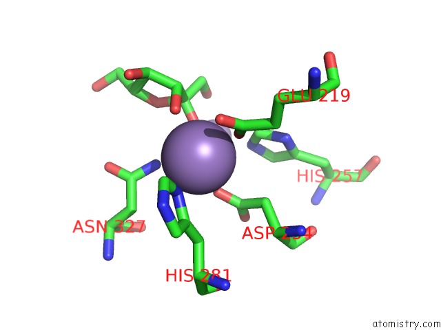
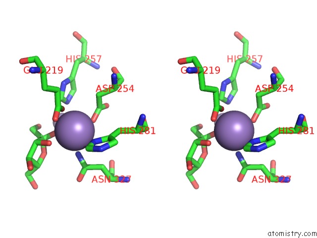
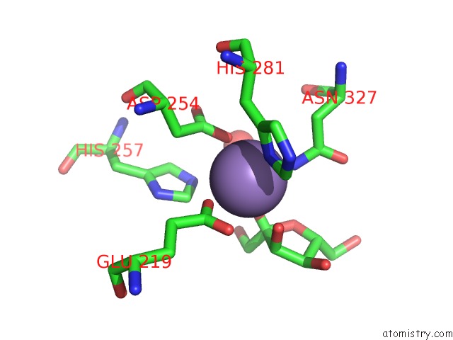
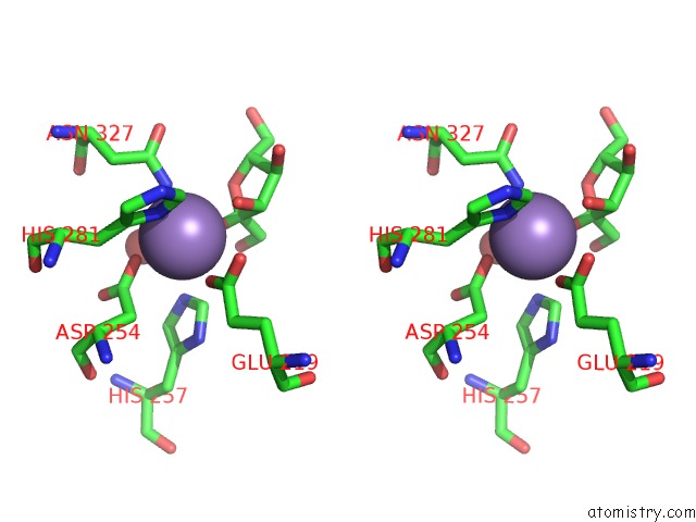
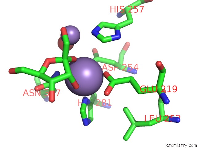
Manganese binding site 1 out of 8 in 3ito
Go back to
Manganese binding site 1 out
of 8 in the Crystal Structure of Pseudomonas Stutzeri L-Rhamnose Isomerase Mutant D327N in Complex with D-Psicose
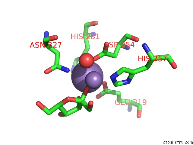
Mono view
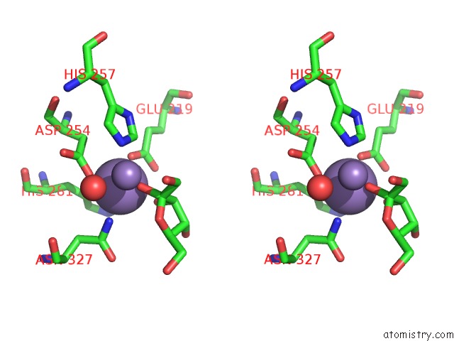
Stereo pair view
Mono view
Stereo pair view
A full contact list of Manganese with other atoms in the Mn binding
site number 1 of Crystal Structure of Pseudomonas Stutzeri L-Rhamnose Isomerase Mutant D327N in Complex with D-Psicose within 5.0Å range:
|
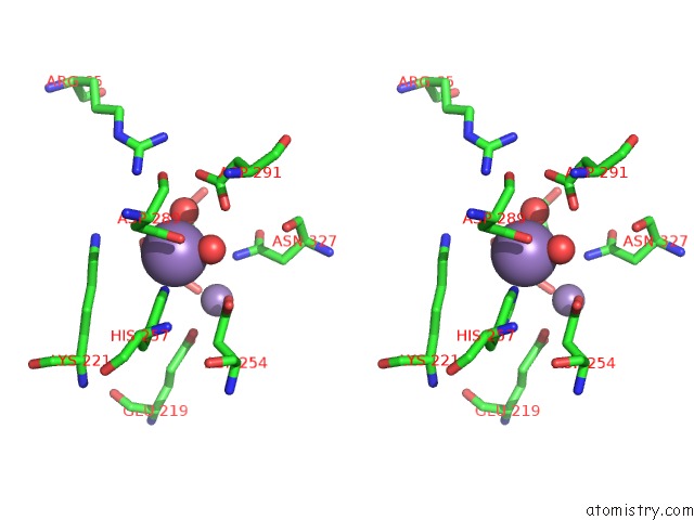
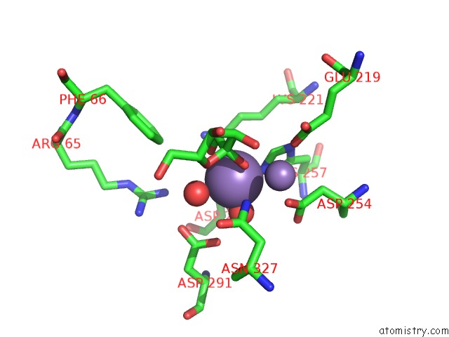
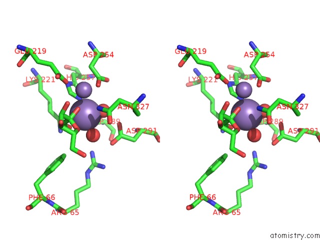
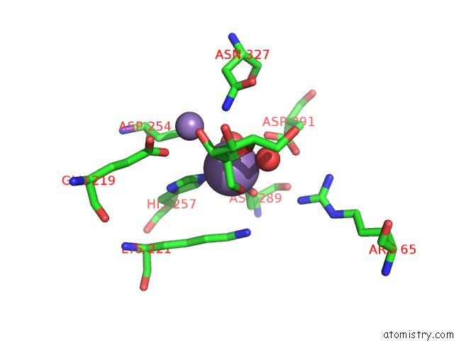
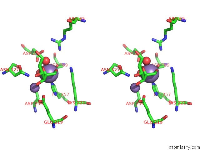
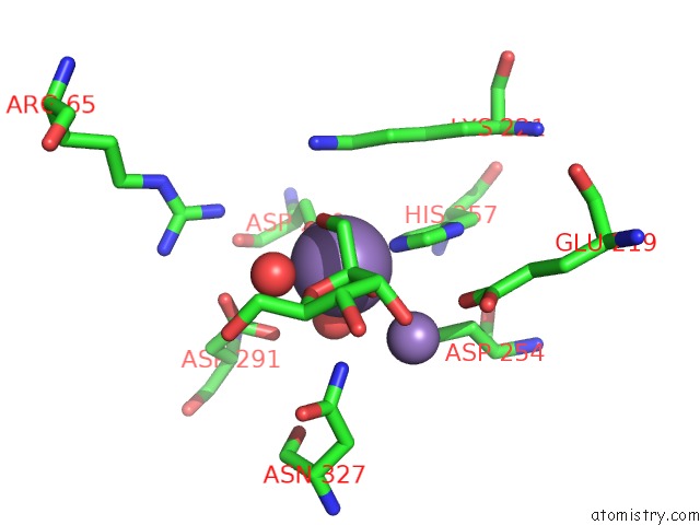
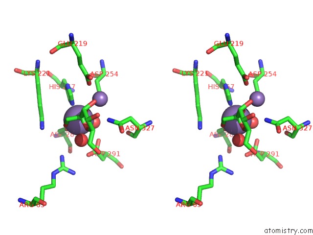
Manganese binding site 2 out of 8 in 3ito
Go back to
Manganese binding site 2 out
of 8 in the Crystal Structure of Pseudomonas Stutzeri L-Rhamnose Isomerase Mutant D327N in Complex with D-Psicose
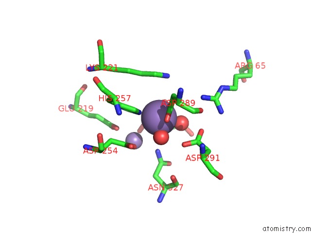
Mono view
Stereo pair view
Mono view
Stereo pair view
A full contact list of Manganese with other atoms in the Mn binding
site number 2 of Crystal Structure of Pseudomonas Stutzeri L-Rhamnose Isomerase Mutant D327N in Complex with D-Psicose within 5.0Å range:
|
Manganese binding site 3 out of 8 in 3ito
Go back to
Manganese binding site 3 out
of 8 in the Crystal Structure of Pseudomonas Stutzeri L-Rhamnose Isomerase Mutant D327N in Complex with D-Psicose
Mono view
Stereo pair view
Mono view
Stereo pair view
A full contact list of Manganese with other atoms in the Mn binding
site number 3 of Crystal Structure of Pseudomonas Stutzeri L-Rhamnose Isomerase Mutant D327N in Complex with D-Psicose within 5.0Å range:
|
Manganese binding site 4 out of 8 in 3ito
Go back to
Manganese binding site 4 out
of 8 in the Crystal Structure of Pseudomonas Stutzeri L-Rhamnose Isomerase Mutant D327N in Complex with D-Psicose
Mono view
Stereo pair view
Mono view
Stereo pair view
A full contact list of Manganese with other atoms in the Mn binding
site number 4 of Crystal Structure of Pseudomonas Stutzeri L-Rhamnose Isomerase Mutant D327N in Complex with D-Psicose within 5.0Å range:
|
Manganese binding site 5 out of 8 in 3ito
Go back to
Manganese binding site 5 out
of 8 in the Crystal Structure of Pseudomonas Stutzeri L-Rhamnose Isomerase Mutant D327N in Complex with D-Psicose
Mono view
Stereo pair view
Mono view
Stereo pair view
A full contact list of Manganese with other atoms in the Mn binding
site number 5 of Crystal Structure of Pseudomonas Stutzeri L-Rhamnose Isomerase Mutant D327N in Complex with D-Psicose within 5.0Å range:
|
Manganese binding site 6 out of 8 in 3ito
Go back to
Manganese binding site 6 out
of 8 in the Crystal Structure of Pseudomonas Stutzeri L-Rhamnose Isomerase Mutant D327N in Complex with D-Psicose
Mono view
Stereo pair view
Mono view
Stereo pair view
A full contact list of Manganese with other atoms in the Mn binding
site number 6 of Crystal Structure of Pseudomonas Stutzeri L-Rhamnose Isomerase Mutant D327N in Complex with D-Psicose within 5.0Å range:
|
Manganese binding site 7 out of 8 in 3ito
Go back to
Manganese binding site 7 out
of 8 in the Crystal Structure of Pseudomonas Stutzeri L-Rhamnose Isomerase Mutant D327N in Complex with D-Psicose
Mono view

Stereo pair view
Mono view
Stereo pair view
A full contact list of Manganese with other atoms in the Mn binding
site number 7 of Crystal Structure of Pseudomonas Stutzeri L-Rhamnose Isomerase Mutant D327N in Complex with D-Psicose within 5.0Å range:
|
Manganese binding site 8 out of 8 in 3ito
Go back to
Manganese binding site 8 out
of 8 in the Crystal Structure of Pseudomonas Stutzeri L-Rhamnose Isomerase Mutant D327N in Complex with D-Psicose
Mono view
Stereo pair view
Mono view
Stereo pair view
A full contact list of Manganese with other atoms in the Mn binding
site number 8 of Crystal Structure of Pseudomonas Stutzeri L-Rhamnose Isomerase Mutant D327N in Complex with D-Psicose within 5.0Å range:
|
Reference:
H.Yoshida,
M.Yamaji,
T.Ishii,
K.Izumori,
S.Kamitori.
Catalytic Reaction Mechanism of Pseudomonas Stutzeri L-Rhamnose Isomerase Deduced From X-Ray Structures Febs J. V. 277 1045 2010.
ISSN: ISSN 1742-464X
PubMed: 20088877
DOI: 10.1111/J.1742-4658.2009.07548.X
Page generated: Sat Oct 5 16:37:39 2024
ISSN: ISSN 1742-464X
PubMed: 20088877
DOI: 10.1111/J.1742-4658.2009.07548.X
Last articles
Zn in 9J0NZn in 9J0O
Zn in 9J0P
Zn in 9FJX
Zn in 9EKB
Zn in 9C0F
Zn in 9CAH
Zn in 9CH0
Zn in 9CH3
Zn in 9CH1