Manganese »
PDB 3hw6-3ki9 »
3ioj »
Manganese in PDB 3ioj: Crystal Structure of the Fucosylgalactoside Alpha N- Acetylgalactosaminyltransferase (Gta, Cisab Mutant L266G, G268A) in Complex with Udp
Enzymatic activity of Crystal Structure of the Fucosylgalactoside Alpha N- Acetylgalactosaminyltransferase (Gta, Cisab Mutant L266G, G268A) in Complex with Udp
All present enzymatic activity of Crystal Structure of the Fucosylgalactoside Alpha N- Acetylgalactosaminyltransferase (Gta, Cisab Mutant L266G, G268A) in Complex with Udp:
2.4.1.37; 2.4.1.40;
2.4.1.37; 2.4.1.40;
Protein crystallography data
The structure of Crystal Structure of the Fucosylgalactoside Alpha N- Acetylgalactosaminyltransferase (Gta, Cisab Mutant L266G, G268A) in Complex with Udp, PDB code: 3ioj
was solved by
T.Pesnot,
R.Jorgensen,
M.M.Palcic,
G.K.Wagner,
with X-Ray Crystallography technique. A brief refinement statistics is given in the table below:
| Resolution Low / High (Å) | 33.13 / 1.65 |
| Space group | P 21 21 21 |
| Cell size a, b, c (Å), α, β, γ (°) | 52.450, 77.536, 153.605, 90.00, 90.00, 90.00 |
| R / Rfree (%) | 16.3 / 19.3 |
Manganese Binding Sites:
The binding sites of Manganese atom in the Crystal Structure of the Fucosylgalactoside Alpha N- Acetylgalactosaminyltransferase (Gta, Cisab Mutant L266G, G268A) in Complex with Udp
(pdb code 3ioj). This binding sites where shown within
5.0 Angstroms radius around Manganese atom.
In total 2 binding sites of Manganese where determined in the Crystal Structure of the Fucosylgalactoside Alpha N- Acetylgalactosaminyltransferase (Gta, Cisab Mutant L266G, G268A) in Complex with Udp, PDB code: 3ioj:
Jump to Manganese binding site number: 1; 2;
In total 2 binding sites of Manganese where determined in the Crystal Structure of the Fucosylgalactoside Alpha N- Acetylgalactosaminyltransferase (Gta, Cisab Mutant L266G, G268A) in Complex with Udp, PDB code: 3ioj:
Jump to Manganese binding site number: 1; 2;
Manganese binding site 1 out of 2 in 3ioj
Go back to
Manganese binding site 1 out
of 2 in the Crystal Structure of the Fucosylgalactoside Alpha N- Acetylgalactosaminyltransferase (Gta, Cisab Mutant L266G, G268A) in Complex with Udp
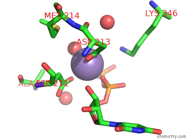
Mono view
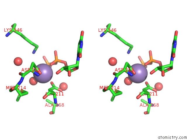
Stereo pair view
Mono view
Stereo pair view
A full contact list of Manganese with other atoms in the Mn binding
site number 1 of Crystal Structure of the Fucosylgalactoside Alpha N- Acetylgalactosaminyltransferase (Gta, Cisab Mutant L266G, G268A) in Complex with Udp within 5.0Å range:
|
Manganese binding site 2 out of 2 in 3ioj
Go back to
Manganese binding site 2 out
of 2 in the Crystal Structure of the Fucosylgalactoside Alpha N- Acetylgalactosaminyltransferase (Gta, Cisab Mutant L266G, G268A) in Complex with Udp
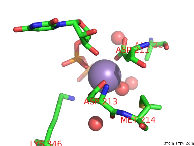
Mono view
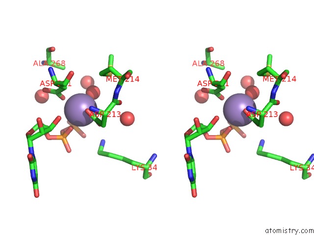
Stereo pair view
Mono view
Stereo pair view
A full contact list of Manganese with other atoms in the Mn binding
site number 2 of Crystal Structure of the Fucosylgalactoside Alpha N- Acetylgalactosaminyltransferase (Gta, Cisab Mutant L266G, G268A) in Complex with Udp within 5.0Å range:
|
Reference:
T.Pesnot,
R.Jorgensen,
M.M.Palcic,
G.K.Wagner.
Structural and Mechanistic Basis For A New Mode of Glycosyltransferase Inhibition. Nat.Chem.Biol. V. 6 321 2010.
ISSN: ISSN 1552-4450
PubMed: 20364127
DOI: 10.1038/NCHEMBIO.343
Page generated: Sat Oct 5 16:35:40 2024
ISSN: ISSN 1552-4450
PubMed: 20364127
DOI: 10.1038/NCHEMBIO.343
Last articles
Zn in 9J0NZn in 9J0O
Zn in 9J0P
Zn in 9FJX
Zn in 9EKB
Zn in 9C0F
Zn in 9CAH
Zn in 9CH0
Zn in 9CH3
Zn in 9CH1