Manganese »
PDB 3hw6-3ki9 »
3i0g »
Manganese in PDB 3i0g: Crystal Structure of Gtb C80S/C196S + Da + Udp-Gal
Enzymatic activity of Crystal Structure of Gtb C80S/C196S + Da + Udp-Gal
All present enzymatic activity of Crystal Structure of Gtb C80S/C196S + Da + Udp-Gal:
2.4.1.37;
2.4.1.37;
Protein crystallography data
The structure of Crystal Structure of Gtb C80S/C196S + Da + Udp-Gal, PDB code: 3i0g
was solved by
B.Schuman,
M.Persson,
R.C.Landry,
R.Polakowski,
J.T.Weadge,
N.O.L.Seto,
S.Borisova,
M.M.Palcic,
S.V.Evans,
with X-Ray Crystallography technique. A brief refinement statistics is given in the table below:
| Resolution Low / High (Å) | 19.87 / 1.40 |
| Space group | C 2 2 21 |
| Cell size a, b, c (Å), α, β, γ (°) | 52.545, 149.277, 79.454, 90.00, 90.00, 90.00 |
| R / Rfree (%) | 19.5 / 23 |
Manganese Binding Sites:
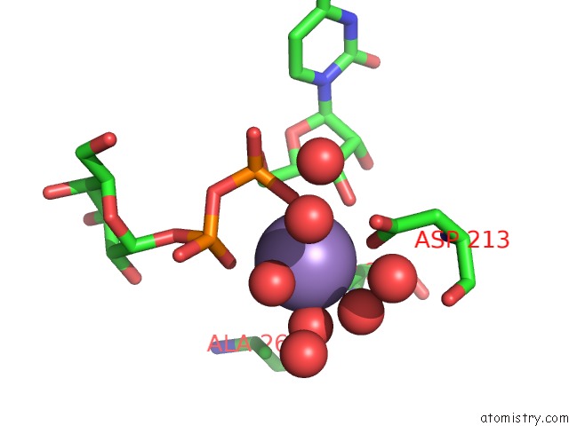
The binding sites of Manganese atom in the Crystal Structure of Gtb C80S/C196S + Da + Udp-Gal
(pdb code 3i0g). This binding sites where shown within
5.0 Angstroms radius around Manganese atom.
In total only one binding site of Manganese was determined in the Crystal Structure of Gtb C80S/C196S + Da + Udp-Gal, PDB code: 3i0g:
In total only one binding site of Manganese was determined in the Crystal Structure of Gtb C80S/C196S + Da + Udp-Gal, PDB code: 3i0g:
Manganese binding site 1 out of 1 in 3i0g
Go back to
Manganese binding site 1 out
of 1 in the Crystal Structure of Gtb C80S/C196S + Da + Udp-Gal
Mono view

Stereo pair view
Mono view
Stereo pair view
A full contact list of Manganese with other atoms in the Mn binding
site number 1 of Crystal Structure of Gtb C80S/C196S + Da + Udp-Gal within 5.0Å range:
|
Reference:
B.Schuman,
M.Persson,
R.C.Landry,
R.Polakowski,
J.T.Weadge,
N.O.Seto,
S.N.Borisova,
M.M.Palcic,
S.V.Evans.
Cysteine-to-Serine Mutants Dramatically Reorder the Active Site of Human Abo(H) Blood Group B Glycosyltransferase Without Affecting Activity: Structural Insights Into Cooperative Substrate Binding J.Mol.Biol. V. 402 399 2010.
ISSN: ISSN 0022-2836
PubMed: 20655926
DOI: 10.1016/J.JMB.2010.07.036
Page generated: Sat Oct 5 16:33:50 2024
ISSN: ISSN 0022-2836
PubMed: 20655926
DOI: 10.1016/J.JMB.2010.07.036
Last articles
Zn in 9J0NZn in 9J0O
Zn in 9J0P
Zn in 9FJX
Zn in 9EKB
Zn in 9C0F
Zn in 9CAH
Zn in 9CH0
Zn in 9CH3
Zn in 9CH1