Manganese »
PDB 3g0z-3hvq »
3hq1 »
Manganese in PDB 3hq1: Crystal Structure of Mycobacterium Tuberculosis Leua Complexed with Citrate and MN2+
Enzymatic activity of Crystal Structure of Mycobacterium Tuberculosis Leua Complexed with Citrate and MN2+
All present enzymatic activity of Crystal Structure of Mycobacterium Tuberculosis Leua Complexed with Citrate and MN2+:
2.3.3.13;
2.3.3.13;
Protein crystallography data
The structure of Crystal Structure of Mycobacterium Tuberculosis Leua Complexed with Citrate and MN2+, PDB code: 3hq1
was solved by
N.Koon,
C.J.Squire,
E.N.Baker,
with X-Ray Crystallography technique. A brief refinement statistics is given in the table below:
| Resolution Low / High (Å) | 38.69 / 1.70 |
| Space group | P 1 21 1 |
| Cell size a, b, c (Å), α, β, γ (°) | 54.361, 154.803, 69.115, 90.00, 98.04, 90.00 |
| R / Rfree (%) | 17 / 20.4 |
Other elements in 3hq1:
The structure of Crystal Structure of Mycobacterium Tuberculosis Leua Complexed with Citrate and MN2+ also contains other interesting chemical elements:
| Chlorine | (Cl) | 2 atoms |
Manganese Binding Sites:
The binding sites of Manganese atom in the Crystal Structure of Mycobacterium Tuberculosis Leua Complexed with Citrate and MN2+
(pdb code 3hq1). This binding sites where shown within
5.0 Angstroms radius around Manganese atom.
In total 3 binding sites of Manganese where determined in the Crystal Structure of Mycobacterium Tuberculosis Leua Complexed with Citrate and MN2+, PDB code: 3hq1:
Jump to Manganese binding site number: 1; 2; 3;
In total 3 binding sites of Manganese where determined in the Crystal Structure of Mycobacterium Tuberculosis Leua Complexed with Citrate and MN2+, PDB code: 3hq1:
Jump to Manganese binding site number: 1; 2; 3;
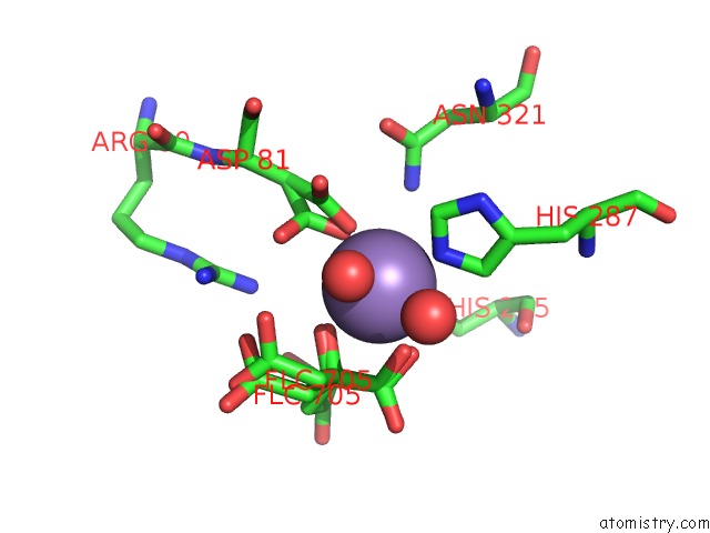
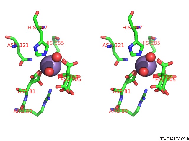
Manganese binding site 1 out of 3 in 3hq1
Go back to
Manganese binding site 1 out
of 3 in the Crystal Structure of Mycobacterium Tuberculosis Leua Complexed with Citrate and MN2+
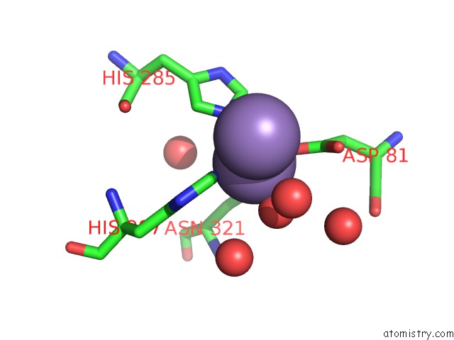
Mono view
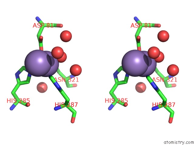
Stereo pair view
Mono view
Stereo pair view
A full contact list of Manganese with other atoms in the Mn binding
site number 1 of Crystal Structure of Mycobacterium Tuberculosis Leua Complexed with Citrate and MN2+ within 5.0Å range:
|
Manganese binding site 2 out of 3 in 3hq1
Go back to
Manganese binding site 2 out
of 3 in the Crystal Structure of Mycobacterium Tuberculosis Leua Complexed with Citrate and MN2+
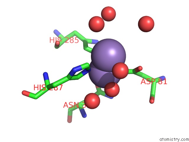
Mono view
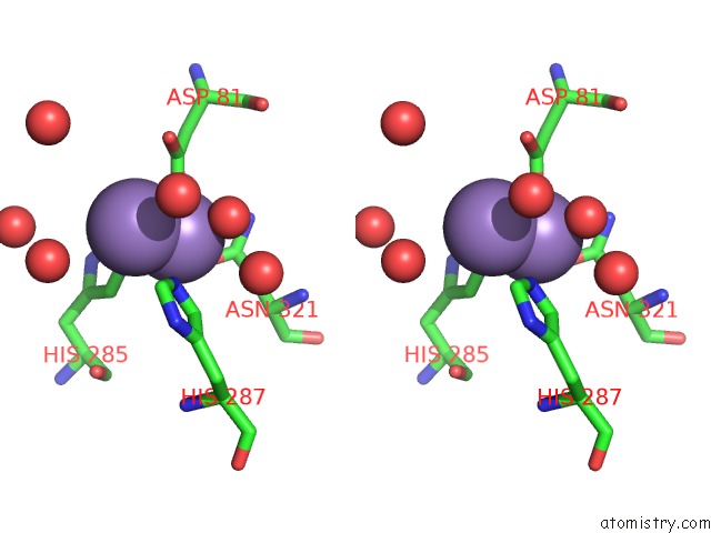
Stereo pair view
Mono view
Stereo pair view
A full contact list of Manganese with other atoms in the Mn binding
site number 2 of Crystal Structure of Mycobacterium Tuberculosis Leua Complexed with Citrate and MN2+ within 5.0Å range:
|
Manganese binding site 3 out of 3 in 3hq1
Go back to
Manganese binding site 3 out
of 3 in the Crystal Structure of Mycobacterium Tuberculosis Leua Complexed with Citrate and MN2+
Mono view
Stereo pair view
Mono view
Stereo pair view
A full contact list of Manganese with other atoms in the Mn binding
site number 3 of Crystal Structure of Mycobacterium Tuberculosis Leua Complexed with Citrate and MN2+ within 5.0Å range:
|
Reference:
N.Koon,
C.J.Squire,
E.N.Baker.
Probing the Active Site of M. Tuberculosis Leua To Be Published.
Page generated: Sat Aug 16 11:55:40 2025
Last articles
Mn in 8DGEMn in 8DJY
Mn in 8DJV
Mn in 8DIP
Mn in 8DHN
Mn in 8DC9
Mn in 8DDE
Mn in 8DDB
Mn in 8DAI
Mn in 8DAL