Manganese »
PDB 3g0z-3hvq »
3ho8 »
Manganese in PDB 3ho8: Crystal Structure of S. Aureus Pyruvate Carboxylase in Complex with Coenzyme A
Protein crystallography data
The structure of Crystal Structure of S. Aureus Pyruvate Carboxylase in Complex with Coenzyme A, PDB code: 3ho8
was solved by
L.Tong,
L.P.C.Yu,
with X-Ray Crystallography technique. A brief refinement statistics is given in the table below:
| Resolution Low / High (Å) | 29.97 / 2.90 |
| Space group | P 21 21 21 |
| Cell size a, b, c (Å), α, β, γ (°) | 96.586, 164.466, 373.346, 90.00, 90.00, 90.00 |
| R / Rfree (%) | 26.4 / 32.8 |
Manganese Binding Sites:
The binding sites of Manganese atom in the Crystal Structure of S. Aureus Pyruvate Carboxylase in Complex with Coenzyme A
(pdb code 3ho8). This binding sites where shown within
5.0 Angstroms radius around Manganese atom.
In total 4 binding sites of Manganese where determined in the Crystal Structure of S. Aureus Pyruvate Carboxylase in Complex with Coenzyme A, PDB code: 3ho8:
Jump to Manganese binding site number: 1; 2; 3; 4;
In total 4 binding sites of Manganese where determined in the Crystal Structure of S. Aureus Pyruvate Carboxylase in Complex with Coenzyme A, PDB code: 3ho8:
Jump to Manganese binding site number: 1; 2; 3; 4;
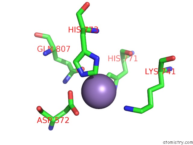
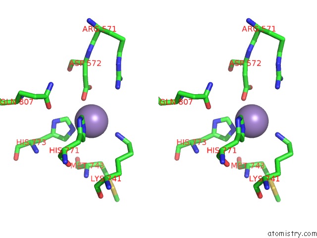
Manganese binding site 1 out of 4 in 3ho8
Go back to
Manganese binding site 1 out
of 4 in the Crystal Structure of S. Aureus Pyruvate Carboxylase in Complex with Coenzyme A
Mono view
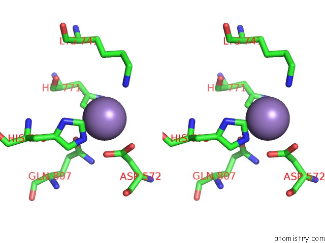
Stereo pair view
Mono view
Stereo pair view
A full contact list of Manganese with other atoms in the Mn binding
site number 1 of Crystal Structure of S. Aureus Pyruvate Carboxylase in Complex with Coenzyme A within 5.0Å range:
|
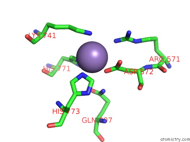
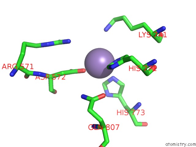
Manganese binding site 2 out of 4 in 3ho8
Go back to
Manganese binding site 2 out
of 4 in the Crystal Structure of S. Aureus Pyruvate Carboxylase in Complex with Coenzyme A
Mono view

Stereo pair view
Mono view
Stereo pair view
A full contact list of Manganese with other atoms in the Mn binding
site number 2 of Crystal Structure of S. Aureus Pyruvate Carboxylase in Complex with Coenzyme A within 5.0Å range:
|
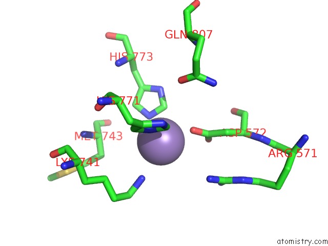
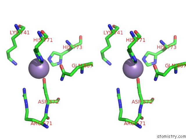
Manganese binding site 3 out of 4 in 3ho8
Go back to
Manganese binding site 3 out
of 4 in the Crystal Structure of S. Aureus Pyruvate Carboxylase in Complex with Coenzyme A
Mono view
Stereo pair view
Mono view
Stereo pair view
A full contact list of Manganese with other atoms in the Mn binding
site number 3 of Crystal Structure of S. Aureus Pyruvate Carboxylase in Complex with Coenzyme A within 5.0Å range:
|
Manganese binding site 4 out of 4 in 3ho8
Go back to
Manganese binding site 4 out
of 4 in the Crystal Structure of S. Aureus Pyruvate Carboxylase in Complex with Coenzyme A
Mono view
Stereo pair view
Mono view
Stereo pair view
A full contact list of Manganese with other atoms in the Mn binding
site number 4 of Crystal Structure of S. Aureus Pyruvate Carboxylase in Complex with Coenzyme A within 5.0Å range:
|
Reference:
L.P.Yu,
S.Xiang,
G.Lasso,
D.Gil,
M.Valle,
L.Tong.
A Symmetrical Tetramer For S. Aureus Pyruvate Carboxylase in Complex with Coenzyme A. Structure V. 17 823 2009.
ISSN: ISSN 0969-2126
PubMed: 19523900
DOI: 10.1016/J.STR.2009.04.008
Page generated: Sat Oct 5 16:31:41 2024
ISSN: ISSN 0969-2126
PubMed: 19523900
DOI: 10.1016/J.STR.2009.04.008
Last articles
Zn in 9J0NZn in 9J0O
Zn in 9J0P
Zn in 9FJX
Zn in 9EKB
Zn in 9C0F
Zn in 9CAH
Zn in 9CH0
Zn in 9CH3
Zn in 9CH1