Manganese »
PDB 3g0z-3hvq »
3h8g »
Manganese in PDB 3h8g: Bestatin Complex Structure of Leucine Aminopeptidase From Pseudomonas Putida
Enzymatic activity of Bestatin Complex Structure of Leucine Aminopeptidase From Pseudomonas Putida
All present enzymatic activity of Bestatin Complex Structure of Leucine Aminopeptidase From Pseudomonas Putida:
3.4.11.1;
3.4.11.1;
Protein crystallography data
The structure of Bestatin Complex Structure of Leucine Aminopeptidase From Pseudomonas Putida, PDB code: 3h8g
was solved by
A.Kale,
B.W.Dijkstra,
T.Sonke,
A.M.W.H.Thunnissen,
with X-Ray Crystallography technique. A brief refinement statistics is given in the table below:
| Resolution Low / High (Å) | 38.63 / 1.50 |
| Space group | P 1 |
| Cell size a, b, c (Å), α, β, γ (°) | 95.976, 95.989, 95.998, 100.82, 107.78, 93.23 |
| R / Rfree (%) | 14.9 / 17.3 |
Other elements in 3h8g:
The structure of Bestatin Complex Structure of Leucine Aminopeptidase From Pseudomonas Putida also contains other interesting chemical elements:
| Potassium | (K) | 6 atoms |
| Zinc | (Zn) | 6 atoms |
Manganese Binding Sites:
The binding sites of Manganese atom in the Bestatin Complex Structure of Leucine Aminopeptidase From Pseudomonas Putida
(pdb code 3h8g). This binding sites where shown within
5.0 Angstroms radius around Manganese atom.
In total 6 binding sites of Manganese where determined in the Bestatin Complex Structure of Leucine Aminopeptidase From Pseudomonas Putida, PDB code: 3h8g:
Jump to Manganese binding site number: 1; 2; 3; 4; 5; 6;
In total 6 binding sites of Manganese where determined in the Bestatin Complex Structure of Leucine Aminopeptidase From Pseudomonas Putida, PDB code: 3h8g:
Jump to Manganese binding site number: 1; 2; 3; 4; 5; 6;
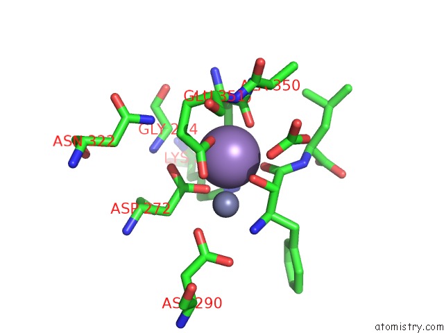
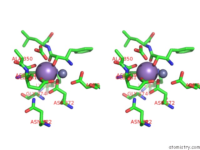
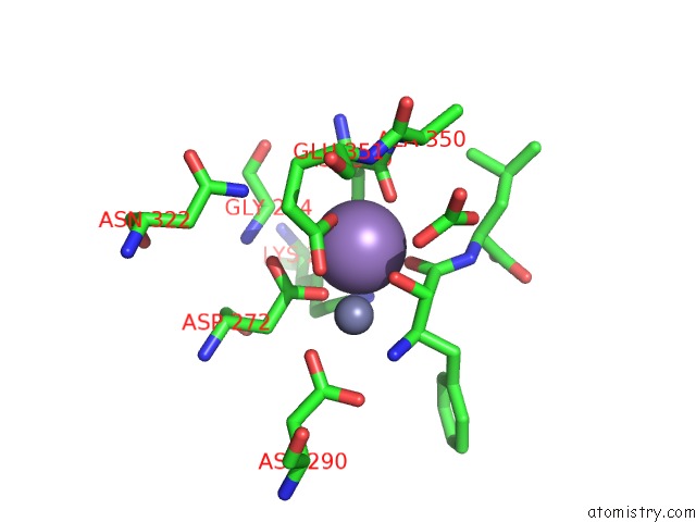
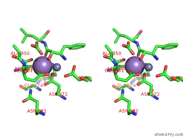
Manganese binding site 1 out of 6 in 3h8g
Go back to
Manganese binding site 1 out
of 6 in the Bestatin Complex Structure of Leucine Aminopeptidase From Pseudomonas Putida
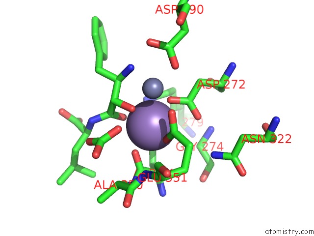
Mono view
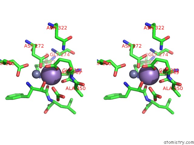
Stereo pair view
Mono view
Stereo pair view
A full contact list of Manganese with other atoms in the Mn binding
site number 1 of Bestatin Complex Structure of Leucine Aminopeptidase From Pseudomonas Putida within 5.0Å range:
|
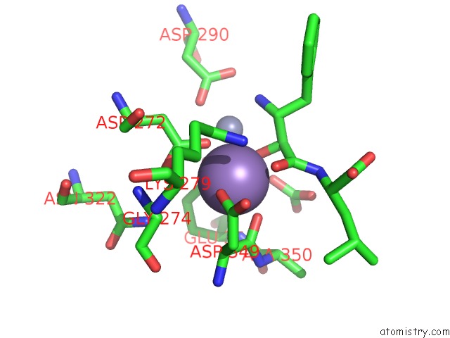
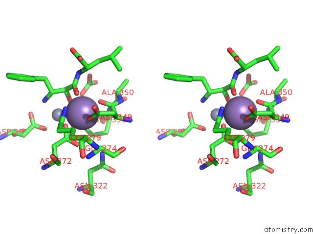
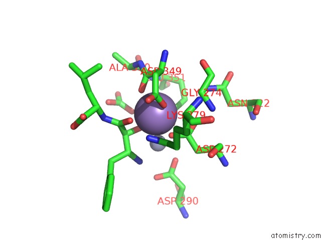
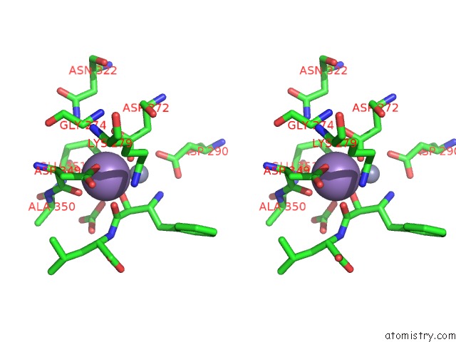
Manganese binding site 2 out of 6 in 3h8g
Go back to
Manganese binding site 2 out
of 6 in the Bestatin Complex Structure of Leucine Aminopeptidase From Pseudomonas Putida
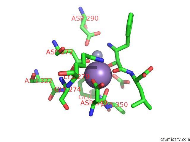
Mono view
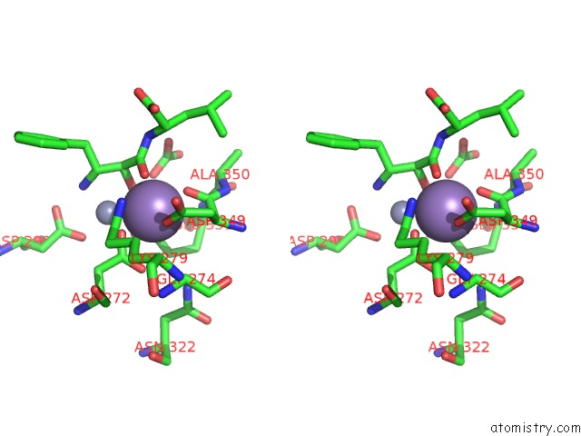
Stereo pair view
Mono view
Stereo pair view
A full contact list of Manganese with other atoms in the Mn binding
site number 2 of Bestatin Complex Structure of Leucine Aminopeptidase From Pseudomonas Putida within 5.0Å range:
|
Manganese binding site 3 out of 6 in 3h8g
Go back to
Manganese binding site 3 out
of 6 in the Bestatin Complex Structure of Leucine Aminopeptidase From Pseudomonas Putida
Mono view
Stereo pair view
Mono view
Stereo pair view
A full contact list of Manganese with other atoms in the Mn binding
site number 3 of Bestatin Complex Structure of Leucine Aminopeptidase From Pseudomonas Putida within 5.0Å range:
|
Manganese binding site 4 out of 6 in 3h8g
Go back to
Manganese binding site 4 out
of 6 in the Bestatin Complex Structure of Leucine Aminopeptidase From Pseudomonas Putida
Mono view
Stereo pair view
Mono view
Stereo pair view
A full contact list of Manganese with other atoms in the Mn binding
site number 4 of Bestatin Complex Structure of Leucine Aminopeptidase From Pseudomonas Putida within 5.0Å range:
|
Manganese binding site 5 out of 6 in 3h8g
Go back to
Manganese binding site 5 out
of 6 in the Bestatin Complex Structure of Leucine Aminopeptidase From Pseudomonas Putida
Mono view
Stereo pair view
Mono view
Stereo pair view
A full contact list of Manganese with other atoms in the Mn binding
site number 5 of Bestatin Complex Structure of Leucine Aminopeptidase From Pseudomonas Putida within 5.0Å range:
|
Manganese binding site 6 out of 6 in 3h8g
Go back to
Manganese binding site 6 out
of 6 in the Bestatin Complex Structure of Leucine Aminopeptidase From Pseudomonas Putida
Mono view
Stereo pair view
Mono view
Stereo pair view
A full contact list of Manganese with other atoms in the Mn binding
site number 6 of Bestatin Complex Structure of Leucine Aminopeptidase From Pseudomonas Putida within 5.0Å range:
|
Reference:
A.Kale,
T.Pijning,
T.Sonke,
B.W.Dijkstra,
A.M.Thunnissen.
Crystal Structure of the Leucine Aminopeptidase From Pseudomonas Putida Reveals the Molecular Basis For Its Enantioselectivity and Broad Substrate Specificity. J.Mol.Biol. V. 398 703 2010.
ISSN: ISSN 0022-2836
PubMed: 20359484
DOI: 10.1016/J.JMB.2010.03.042
Page generated: Sat Oct 5 16:29:17 2024
ISSN: ISSN 0022-2836
PubMed: 20359484
DOI: 10.1016/J.JMB.2010.03.042
Last articles
Cl in 3DP2Cl in 3DQT
Cl in 3DQS
Cl in 3DP1
Cl in 3DOY
Cl in 3DP0
Cl in 3DOZ
Cl in 3DOL
Cl in 3DOK
Cl in 3DOJ