Manganese »
PDB 3ebc-3g0a »
3fjq »
Manganese in PDB 3fjq: Crystal Structure of Camp-Dependent Protein Kinase Catalytic Subunit Alpha in Complex with Peptide Inhibitor Pki Alpha (6-25)
Enzymatic activity of Crystal Structure of Camp-Dependent Protein Kinase Catalytic Subunit Alpha in Complex with Peptide Inhibitor Pki Alpha (6-25)
All present enzymatic activity of Crystal Structure of Camp-Dependent Protein Kinase Catalytic Subunit Alpha in Complex with Peptide Inhibitor Pki Alpha (6-25):
2.7.11.11;
2.7.11.11;
Protein crystallography data
The structure of Crystal Structure of Camp-Dependent Protein Kinase Catalytic Subunit Alpha in Complex with Peptide Inhibitor Pki Alpha (6-25), PDB code: 3fjq
was solved by
C.Kim,
with X-Ray Crystallography technique. A brief refinement statistics is given in the table below:
| Resolution Low / High (Å) | 50.00 / 1.60 |
| Space group | P 21 21 21 |
| Cell size a, b, c (Å), α, β, γ (°) | 57.517, 80.548, 97.622, 90.00, 90.00, 90.00 |
| R / Rfree (%) | 17.7 / 20.5 |
Manganese Binding Sites:
The binding sites of Manganese atom in the Crystal Structure of Camp-Dependent Protein Kinase Catalytic Subunit Alpha in Complex with Peptide Inhibitor Pki Alpha (6-25)
(pdb code 3fjq). This binding sites where shown within
5.0 Angstroms radius around Manganese atom.
In total 2 binding sites of Manganese where determined in the Crystal Structure of Camp-Dependent Protein Kinase Catalytic Subunit Alpha in Complex with Peptide Inhibitor Pki Alpha (6-25), PDB code: 3fjq:
Jump to Manganese binding site number: 1; 2;
In total 2 binding sites of Manganese where determined in the Crystal Structure of Camp-Dependent Protein Kinase Catalytic Subunit Alpha in Complex with Peptide Inhibitor Pki Alpha (6-25), PDB code: 3fjq:
Jump to Manganese binding site number: 1; 2;
Manganese binding site 1 out of 2 in 3fjq
Go back to
Manganese binding site 1 out
of 2 in the Crystal Structure of Camp-Dependent Protein Kinase Catalytic Subunit Alpha in Complex with Peptide Inhibitor Pki Alpha (6-25)
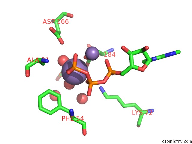
Mono view
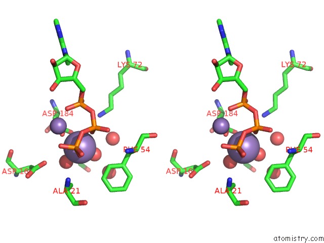
Stereo pair view
Mono view
Stereo pair view
A full contact list of Manganese with other atoms in the Mn binding
site number 1 of Crystal Structure of Camp-Dependent Protein Kinase Catalytic Subunit Alpha in Complex with Peptide Inhibitor Pki Alpha (6-25) within 5.0Å range:
|
Manganese binding site 2 out of 2 in 3fjq
Go back to
Manganese binding site 2 out
of 2 in the Crystal Structure of Camp-Dependent Protein Kinase Catalytic Subunit Alpha in Complex with Peptide Inhibitor Pki Alpha (6-25)
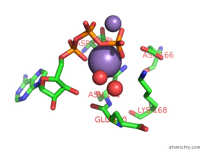
Mono view
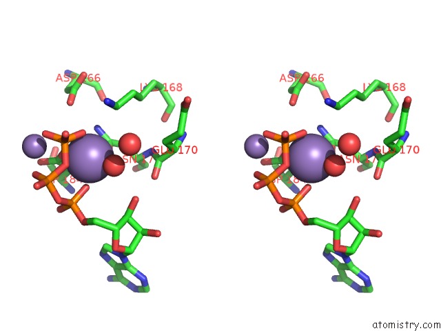
Stereo pair view
Mono view
Stereo pair view
A full contact list of Manganese with other atoms in the Mn binding
site number 2 of Crystal Structure of Camp-Dependent Protein Kinase Catalytic Subunit Alpha in Complex with Peptide Inhibitor Pki Alpha (6-25) within 5.0Å range:
|
Reference:
E.E.Thompson,
A.P.Kornev,
N.Kannan,
C.Kim,
L.F.Ten Eyck,
S.S.Taylor.
Comparative Surface Geometry of the Protein Kinase Family. Protein Sci. V. 18 2016 2009.
ISSN: ISSN 0961-8368
PubMed: 19610074
DOI: 10.1002/PRO.209
Page generated: Sat Oct 5 16:18:10 2024
ISSN: ISSN 0961-8368
PubMed: 19610074
DOI: 10.1002/PRO.209
Last articles
Fe in 7R2SFe in 7R0W
Fe in 7R2R
Fe in 7R2P
Fe in 7R2O
Fe in 7R1J
Fe in 7R1I
Fe in 7QWT
Fe in 7R1H
Fe in 7R0F