Manganese »
PDB 3cev-3ea3 »
3e8z »
Manganese in PDB 3e8z: X-Ray Structure of Rat Arginase I-N130A Mutant: the Unliganded Complex
Enzymatic activity of X-Ray Structure of Rat Arginase I-N130A Mutant: the Unliganded Complex
All present enzymatic activity of X-Ray Structure of Rat Arginase I-N130A Mutant: the Unliganded Complex:
3.5.3.1;
3.5.3.1;
Protein crystallography data
The structure of X-Ray Structure of Rat Arginase I-N130A Mutant: the Unliganded Complex, PDB code: 3e8z
was solved by
E.Y.Shishova,
L.Di Costanzo,
D.W.Christianson,
with X-Ray Crystallography technique. A brief refinement statistics is given in the table below:
| Resolution Low / High (Å) | 41.77 / 2.00 |
| Space group | P 32 |
| Cell size a, b, c (Å), α, β, γ (°) | 87.500, 87.500, 100.110, 90.00, 90.00, 120.00 |
| R / Rfree (%) | 23.6 / 28 |
Manganese Binding Sites:
The binding sites of Manganese atom in the X-Ray Structure of Rat Arginase I-N130A Mutant: the Unliganded Complex
(pdb code 3e8z). This binding sites where shown within
5.0 Angstroms radius around Manganese atom.
In total 6 binding sites of Manganese where determined in the X-Ray Structure of Rat Arginase I-N130A Mutant: the Unliganded Complex, PDB code: 3e8z:
Jump to Manganese binding site number: 1; 2; 3; 4; 5; 6;
In total 6 binding sites of Manganese where determined in the X-Ray Structure of Rat Arginase I-N130A Mutant: the Unliganded Complex, PDB code: 3e8z:
Jump to Manganese binding site number: 1; 2; 3; 4; 5; 6;
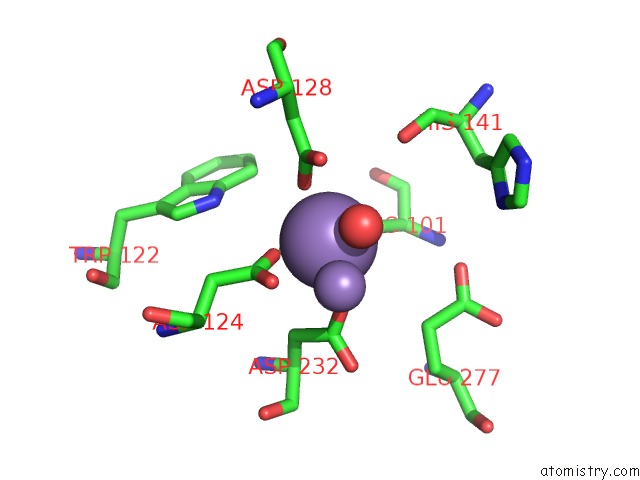
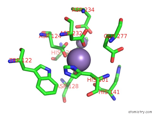
Manganese binding site 1 out of 6 in 3e8z
Go back to
Manganese binding site 1 out
of 6 in the X-Ray Structure of Rat Arginase I-N130A Mutant: the Unliganded Complex
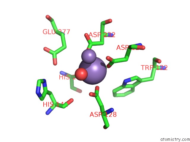
Mono view
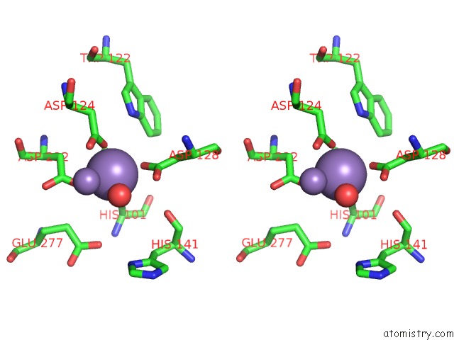
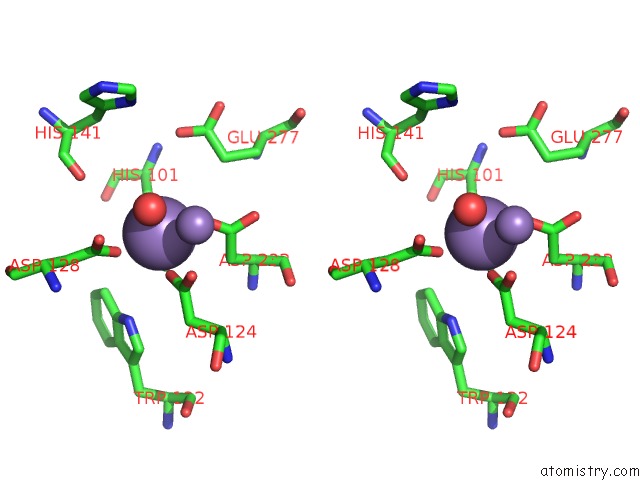
Stereo pair view
Mono view
Stereo pair view
A full contact list of Manganese with other atoms in the Mn binding
site number 1 of X-Ray Structure of Rat Arginase I-N130A Mutant: the Unliganded Complex within 5.0Å range:
|
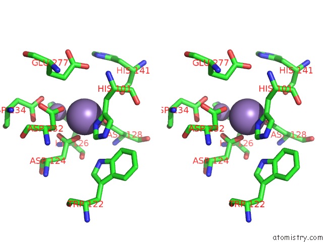
Manganese binding site 2 out of 6 in 3e8z
Go back to
Manganese binding site 2 out
of 6 in the X-Ray Structure of Rat Arginase I-N130A Mutant: the Unliganded Complex
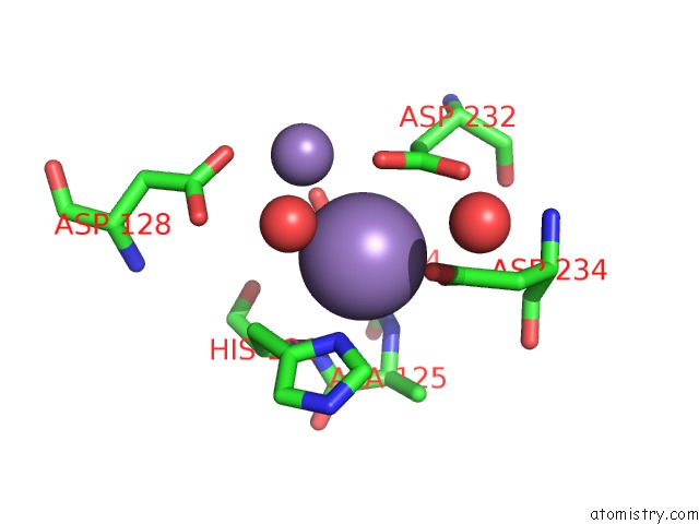
Mono view
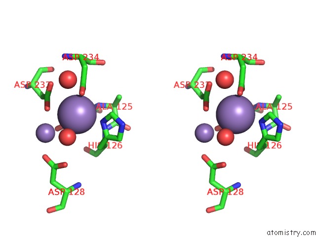
Stereo pair view
Mono view
Stereo pair view
A full contact list of Manganese with other atoms in the Mn binding
site number 2 of X-Ray Structure of Rat Arginase I-N130A Mutant: the Unliganded Complex within 5.0Å range:
|
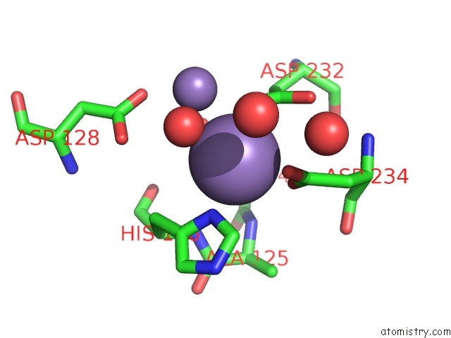
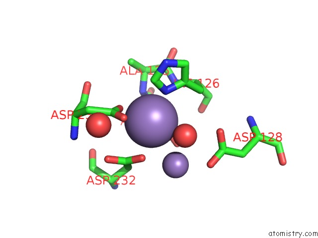
Manganese binding site 3 out of 6 in 3e8z
Go back to
Manganese binding site 3 out
of 6 in the X-Ray Structure of Rat Arginase I-N130A Mutant: the Unliganded Complex
Mono view
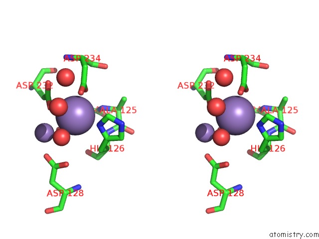
Stereo pair view
Mono view
Stereo pair view
A full contact list of Manganese with other atoms in the Mn binding
site number 3 of X-Ray Structure of Rat Arginase I-N130A Mutant: the Unliganded Complex within 5.0Å range:
|
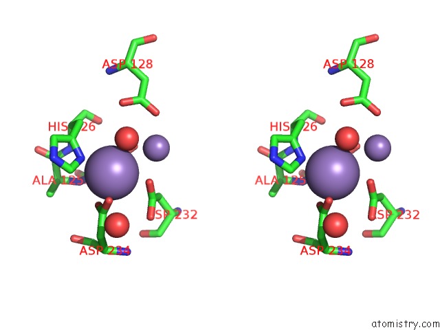
Manganese binding site 4 out of 6 in 3e8z
Go back to
Manganese binding site 4 out
of 6 in the X-Ray Structure of Rat Arginase I-N130A Mutant: the Unliganded Complex
Mono view
Stereo pair view
Mono view
Stereo pair view
A full contact list of Manganese with other atoms in the Mn binding
site number 4 of X-Ray Structure of Rat Arginase I-N130A Mutant: the Unliganded Complex within 5.0Å range:
|
Manganese binding site 5 out of 6 in 3e8z
Go back to
Manganese binding site 5 out
of 6 in the X-Ray Structure of Rat Arginase I-N130A Mutant: the Unliganded Complex
Mono view
Stereo pair view
Mono view
Stereo pair view
A full contact list of Manganese with other atoms in the Mn binding
site number 5 of X-Ray Structure of Rat Arginase I-N130A Mutant: the Unliganded Complex within 5.0Å range:
|
Manganese binding site 6 out of 6 in 3e8z
Go back to
Manganese binding site 6 out
of 6 in the X-Ray Structure of Rat Arginase I-N130A Mutant: the Unliganded Complex
Mono view
Stereo pair view
Mono view
Stereo pair view
A full contact list of Manganese with other atoms in the Mn binding
site number 6 of X-Ray Structure of Rat Arginase I-N130A Mutant: the Unliganded Complex within 5.0Å range:
|
Reference:
E.Y.Shishova,
L.Di Costanzo,
F.A.Emig,
D.E.Ash,
D.W.Christianson.
Probing the Specificity Determinants of Amino Acid Recognition By Arginase. Biochemistry V. 48 121 2009.
ISSN: ISSN 0006-2960
PubMed: 19093830
DOI: 10.1021/BI801911V
Page generated: Sat Oct 5 16:10:11 2024
ISSN: ISSN 0006-2960
PubMed: 19093830
DOI: 10.1021/BI801911V
Last articles
Ca in 5W0RCa in 5VYG
Ca in 5VT8
Ca in 5VYF
Ca in 5VYB
Ca in 5VXZ
Ca in 5VTM
Ca in 5VWM
Ca in 5VTD
Ca in 5VUG