Manganese »
PDB 3cev-3ea3 »
3dky »
Manganese in PDB 3dky: Crystal Structure of the Replication Initiator Protein Encoded on Plasmid PMV158 (Repb), Tetragonal Form, to 3.6 Ang Resolution
Protein crystallography data
The structure of Crystal Structure of the Replication Initiator Protein Encoded on Plasmid PMV158 (Repb), Tetragonal Form, to 3.6 Ang Resolution, PDB code: 3dky
was solved by
D.R.Boer,
J.A.Ruiz-Maso,
A.G.Blanco,
M.Vives-Llacer,
I.Uson,
F.X.Gomis-Ruth,
M.Espinosa,
G.Del Solar,
M.Coll,
with X-Ray Crystallography technique. A brief refinement statistics is given in the table below:
| Resolution Low / High (Å) | 19.57 / 3.60 |
| Space group | P 43 |
| Cell size a, b, c (Å), α, β, γ (°) | 91.460, 91.460, 227.080, 90.00, 90.00, 90.00 |
| R / Rfree (%) | 22.3 / 25.7 |
Manganese Binding Sites:
The binding sites of Manganese atom in the Crystal Structure of the Replication Initiator Protein Encoded on Plasmid PMV158 (Repb), Tetragonal Form, to 3.6 Ang Resolution
(pdb code 3dky). This binding sites where shown within
5.0 Angstroms radius around Manganese atom.
In total 4 binding sites of Manganese where determined in the Crystal Structure of the Replication Initiator Protein Encoded on Plasmid PMV158 (Repb), Tetragonal Form, to 3.6 Ang Resolution, PDB code: 3dky:
Jump to Manganese binding site number: 1; 2; 3; 4;
In total 4 binding sites of Manganese where determined in the Crystal Structure of the Replication Initiator Protein Encoded on Plasmid PMV158 (Repb), Tetragonal Form, to 3.6 Ang Resolution, PDB code: 3dky:
Jump to Manganese binding site number: 1; 2; 3; 4;
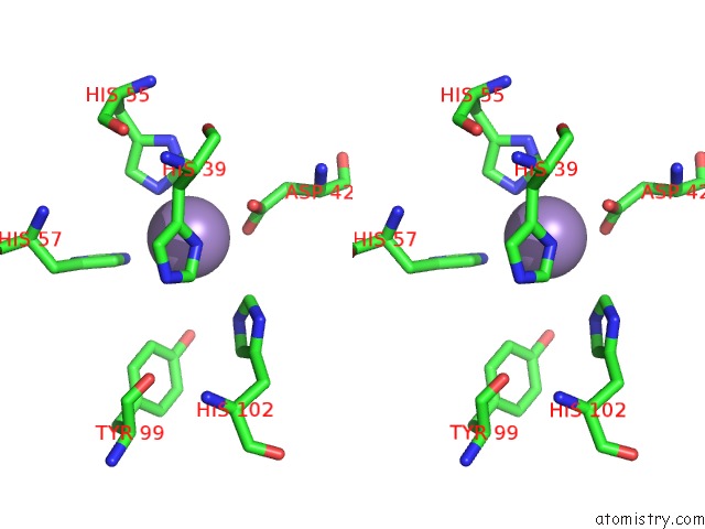
Manganese binding site 1 out of 4 in 3dky
Go back to
Manganese binding site 1 out
of 4 in the Crystal Structure of the Replication Initiator Protein Encoded on Plasmid PMV158 (Repb), Tetragonal Form, to 3.6 Ang Resolution
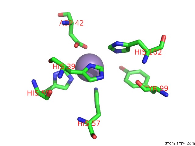
Mono view
Stereo pair view
Mono view
Stereo pair view
A full contact list of Manganese with other atoms in the Mn binding
site number 1 of Crystal Structure of the Replication Initiator Protein Encoded on Plasmid PMV158 (Repb), Tetragonal Form, to 3.6 Ang Resolution within 5.0Å range:
|
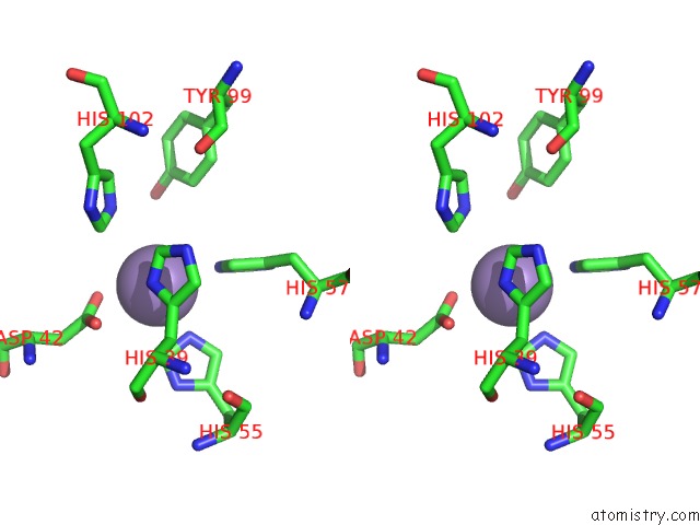
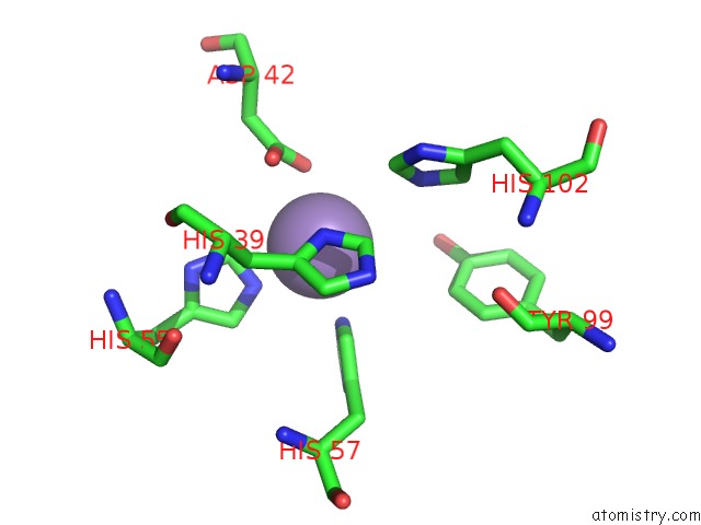
Manganese binding site 2 out of 4 in 3dky
Go back to
Manganese binding site 2 out
of 4 in the Crystal Structure of the Replication Initiator Protein Encoded on Plasmid PMV158 (Repb), Tetragonal Form, to 3.6 Ang Resolution
Mono view
Stereo pair view
Mono view
Stereo pair view
A full contact list of Manganese with other atoms in the Mn binding
site number 2 of Crystal Structure of the Replication Initiator Protein Encoded on Plasmid PMV158 (Repb), Tetragonal Form, to 3.6 Ang Resolution within 5.0Å range:
|
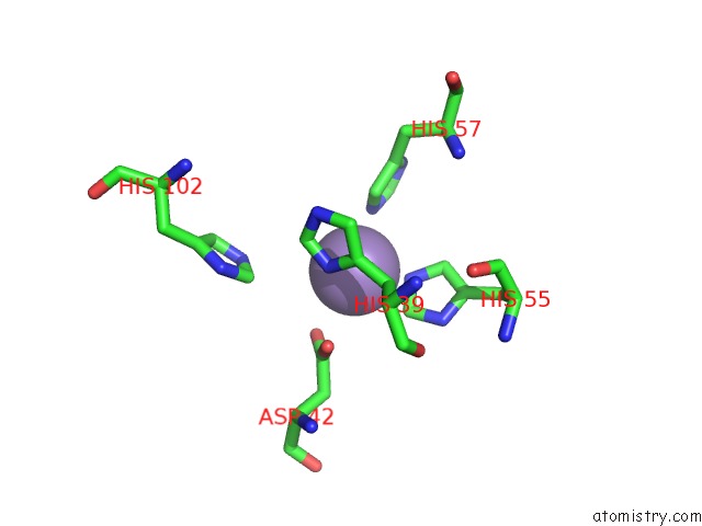
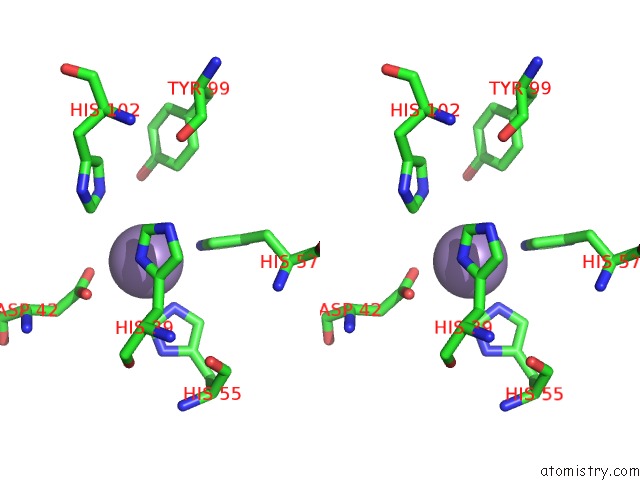
Manganese binding site 3 out of 4 in 3dky
Go back to
Manganese binding site 3 out
of 4 in the Crystal Structure of the Replication Initiator Protein Encoded on Plasmid PMV158 (Repb), Tetragonal Form, to 3.6 Ang Resolution

Mono view
Stereo pair view
Mono view
Stereo pair view
A full contact list of Manganese with other atoms in the Mn binding
site number 3 of Crystal Structure of the Replication Initiator Protein Encoded on Plasmid PMV158 (Repb), Tetragonal Form, to 3.6 Ang Resolution within 5.0Å range:
|
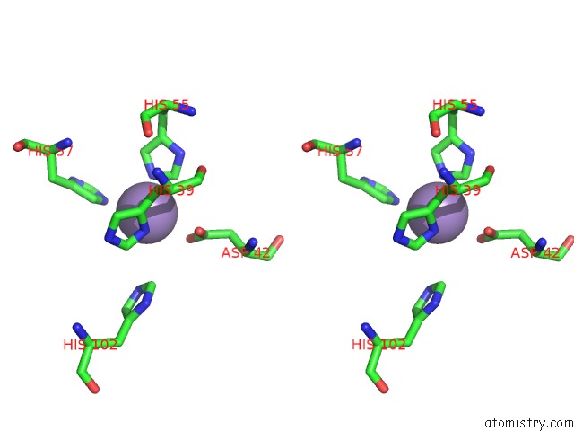
Manganese binding site 4 out of 4 in 3dky
Go back to
Manganese binding site 4 out
of 4 in the Crystal Structure of the Replication Initiator Protein Encoded on Plasmid PMV158 (Repb), Tetragonal Form, to 3.6 Ang Resolution
Mono view
Stereo pair view
Mono view
Stereo pair view
A full contact list of Manganese with other atoms in the Mn binding
site number 4 of Crystal Structure of the Replication Initiator Protein Encoded on Plasmid PMV158 (Repb), Tetragonal Form, to 3.6 Ang Resolution within 5.0Å range:
|
Reference:
D.R.Boer,
J.A.Ruiz-Maso,
J.R.Lopez-Blanco,
A.G.Blanco,
M.Vives-Llacer,
P.Chacon,
I.Uson,
F.X.Gomis-Ruth,
M.Espinosa,
O.Llorca,
G.Del Solar,
M.Coll.
Plasmid Replication Initiator Repb Forms A Hexamer Reminiscent of Ring Helicases and Has Mobile Nuclease Domains Embo J. V. 28 1666 2009.
ISSN: ISSN 0261-4189
PubMed: 19440202
DOI: 10.1038/EMBOJ.2009.125
Page generated: Sat Oct 5 16:05:06 2024
ISSN: ISSN 0261-4189
PubMed: 19440202
DOI: 10.1038/EMBOJ.2009.125
Last articles
Ca in 5S5UCa in 5S5T
Ca in 5S5S
Ca in 5S5R
Ca in 5S5Q
Ca in 5S5P
Ca in 5S5O
Ca in 5S5N
Ca in 5S5M
Ca in 5S5L