Manganese »
PDB 3cev-3ea3 »
3cqw »
Manganese in PDB 3cqw: Crystal Structure of Akt-1 Complexed with Substrate Peptide and Inhibitor
Enzymatic activity of Crystal Structure of Akt-1 Complexed with Substrate Peptide and Inhibitor
All present enzymatic activity of Crystal Structure of Akt-1 Complexed with Substrate Peptide and Inhibitor:
2.7.11.1; 2.7.11.26;
2.7.11.1; 2.7.11.26;
Protein crystallography data
The structure of Crystal Structure of Akt-1 Complexed with Substrate Peptide and Inhibitor, PDB code: 3cqw
was solved by
J.Pandit,
with X-Ray Crystallography technique. A brief refinement statistics is given in the table below:
| Resolution Low / High (Å) | 45.36 / 2.00 |
| Space group | P 1 21 1 |
| Cell size a, b, c (Å), α, β, γ (°) | 42.505, 55.879, 92.864, 90.00, 102.36, 90.00 |
| R / Rfree (%) | 20 / 25.7 |
Other elements in 3cqw:
The structure of Crystal Structure of Akt-1 Complexed with Substrate Peptide and Inhibitor also contains other interesting chemical elements:
| Chlorine | (Cl) | 1 atom |
Manganese Binding Sites:
The binding sites of Manganese atom in the Crystal Structure of Akt-1 Complexed with Substrate Peptide and Inhibitor
(pdb code 3cqw). This binding sites where shown within
5.0 Angstroms radius around Manganese atom.
In total only one binding site of Manganese was determined in the Crystal Structure of Akt-1 Complexed with Substrate Peptide and Inhibitor, PDB code: 3cqw:
In total only one binding site of Manganese was determined in the Crystal Structure of Akt-1 Complexed with Substrate Peptide and Inhibitor, PDB code: 3cqw:
Manganese binding site 1 out of 1 in 3cqw
Go back to
Manganese binding site 1 out
of 1 in the Crystal Structure of Akt-1 Complexed with Substrate Peptide and Inhibitor
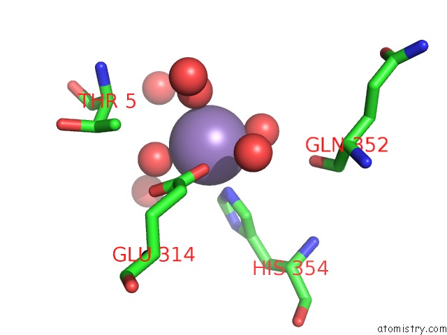
Mono view
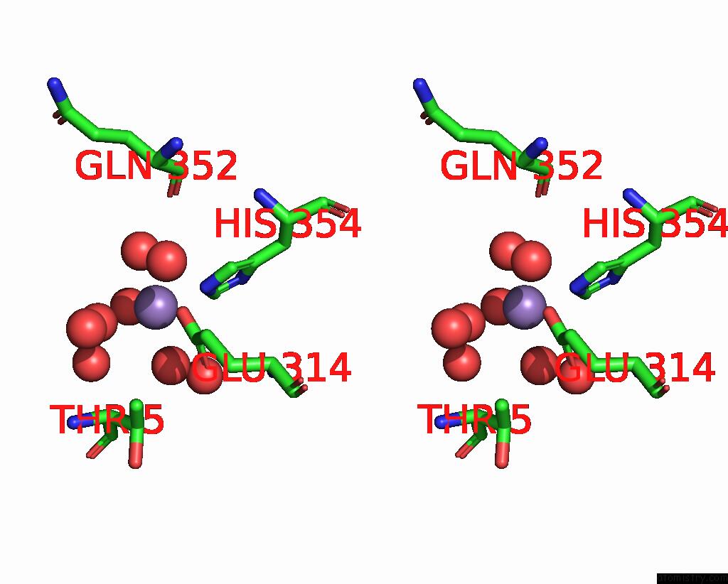
Stereo pair view
Mono view
Stereo pair view
A full contact list of Manganese with other atoms in the Mn binding
site number 1 of Crystal Structure of Akt-1 Complexed with Substrate Peptide and Inhibitor within 5.0Å range:
|
Reference:
B.Lippa,
G.Pan,
M.Corbett,
C.Li,
G.S.Kauffman,
J.Pandit,
S.Robinson,
L.Wei,
E.Kozina,
E.S.Marr,
G.Borzillo,
E.Knauth,
E.G.Barbacci-Tobin,
P.Vincent,
M.Troutman,
D.Baker,
F.Rajamohan,
S.Kakar,
T.Clark,
J.Morris.
Synthesis and Structure Based Optimization of Novel Akt Inhibitors Bioorg.Med.Chem.Lett. V. 18 3359 2008.
ISSN: ISSN 0960-894X
PubMed: 18456494
DOI: 10.1016/J.BMCL.2008.04.034
Page generated: Sat Oct 5 16:02:29 2024
ISSN: ISSN 0960-894X
PubMed: 18456494
DOI: 10.1016/J.BMCL.2008.04.034
Last articles
Zn in 9MJ5Zn in 9HNW
Zn in 9G0L
Zn in 9FNE
Zn in 9DZN
Zn in 9E0I
Zn in 9D32
Zn in 9DAK
Zn in 8ZXC
Zn in 8ZUF