Manganese »
PDB 3cev-3ea3 »
3ckn »
Manganese in PDB 3ckn: Crystal Structure of A Mycobacterial Protein
Protein crystallography data
The structure of Crystal Structure of A Mycobacterial Protein, PDB code: 3ckn
was solved by
Z.Marland,
J.Rossjohn,
with X-Ray Crystallography technique. A brief refinement statistics is given in the table below:
| Resolution Low / High (Å) | 50.00 / 2.20 |
| Space group | P 41 21 2 |
| Cell size a, b, c (Å), α, β, γ (°) | 86.862, 86.862, 104.243, 90.00, 90.00, 90.00 |
| R / Rfree (%) | 20.6 / 23.2 |
Manganese Binding Sites:
The binding sites of Manganese atom in the Crystal Structure of A Mycobacterial Protein
(pdb code 3ckn). This binding sites where shown within
5.0 Angstroms radius around Manganese atom.
In total only one binding site of Manganese was determined in the Crystal Structure of A Mycobacterial Protein, PDB code: 3ckn:
In total only one binding site of Manganese was determined in the Crystal Structure of A Mycobacterial Protein, PDB code: 3ckn:
Manganese binding site 1 out of 1 in 3ckn
Go back to
Manganese binding site 1 out
of 1 in the Crystal Structure of A Mycobacterial Protein
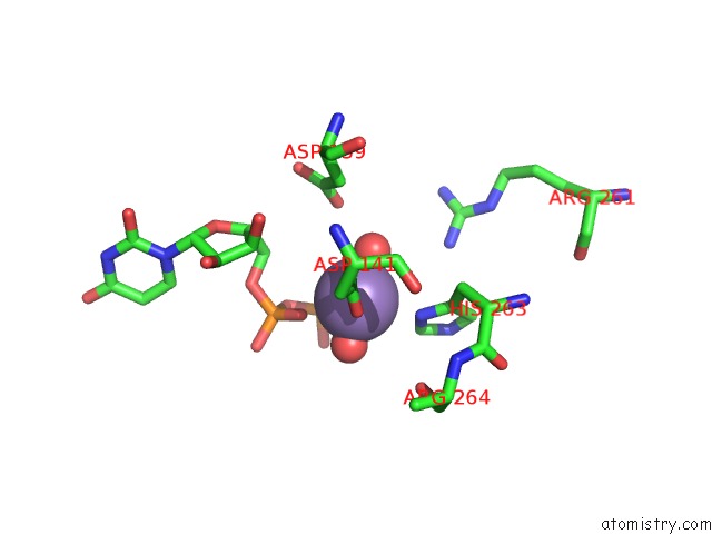
Mono view
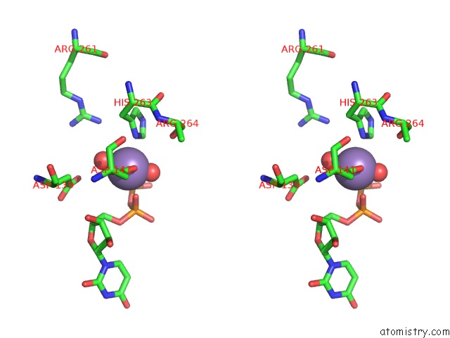
Stereo pair view
Mono view
Stereo pair view
A full contact list of Manganese with other atoms in the Mn binding
site number 1 of Crystal Structure of A Mycobacterial Protein within 5.0Å range:
|
Reference:
Z.Fulton,
A.Mcalister,
M.C.Wilce,
R.Brammananth,
L.Zaker-Tabrizi,
M.A.Perugini,
S.P.Bottomley,
R.L.Coppel,
P.K.Crellin,
J.Rossjohn,
T.Beddoe.
Crystal Structure of A Udp-Glucose-Specific Glycosyltransferase From A Mycobacterium Species. J.Biol.Chem. V. 283 27881 2008.
ISSN: ISSN 0021-9258
PubMed: 18667419
DOI: 10.1074/JBC.M801853200
Page generated: Sat Oct 5 16:02:29 2024
ISSN: ISSN 0021-9258
PubMed: 18667419
DOI: 10.1074/JBC.M801853200
Last articles
Ca in 5NGQCa in 5NH5
Ca in 5NGY
Ca in 5NG1
Ca in 5NFZ
Ca in 5NF3
Ca in 5NF2
Ca in 5NF0
Ca in 5NEY
Ca in 5NES