Manganese »
PDB 3auz-3c5m »
3bza »
Manganese in PDB 3bza: Structure of Mn-Substituted Homoprotocatechuate 2,3-Dioxygenase From B.Fuscum at 1.7 Ang Resolution
Enzymatic activity of Structure of Mn-Substituted Homoprotocatechuate 2,3-Dioxygenase From B.Fuscum at 1.7 Ang Resolution
All present enzymatic activity of Structure of Mn-Substituted Homoprotocatechuate 2,3-Dioxygenase From B.Fuscum at 1.7 Ang Resolution:
1.13.11.15;
1.13.11.15;
Protein crystallography data
The structure of Structure of Mn-Substituted Homoprotocatechuate 2,3-Dioxygenase From B.Fuscum at 1.7 Ang Resolution, PDB code: 3bza
was solved by
E.G.Kovaleva,
J.D.Lipscomb,
with X-Ray Crystallography technique. A brief refinement statistics is given in the table below:
| Resolution Low / High (Å) | 28.82 / 1.70 |
| Space group | P 21 21 2 |
| Cell size a, b, c (Å), α, β, γ (°) | 110.502, 152.191, 96.278, 90.00, 90.00, 90.00 |
| R / Rfree (%) | 17 / 19.7 |
Other elements in 3bza:
The structure of Structure of Mn-Substituted Homoprotocatechuate 2,3-Dioxygenase From B.Fuscum at 1.7 Ang Resolution also contains other interesting chemical elements:
| Chlorine | (Cl) | 4 atoms |
| Calcium | (Ca) | 1 atom |
Manganese Binding Sites:
The binding sites of Manganese atom in the Structure of Mn-Substituted Homoprotocatechuate 2,3-Dioxygenase From B.Fuscum at 1.7 Ang Resolution
(pdb code 3bza). This binding sites where shown within
5.0 Angstroms radius around Manganese atom.
In total 4 binding sites of Manganese where determined in the Structure of Mn-Substituted Homoprotocatechuate 2,3-Dioxygenase From B.Fuscum at 1.7 Ang Resolution, PDB code: 3bza:
Jump to Manganese binding site number: 1; 2; 3; 4;
In total 4 binding sites of Manganese where determined in the Structure of Mn-Substituted Homoprotocatechuate 2,3-Dioxygenase From B.Fuscum at 1.7 Ang Resolution, PDB code: 3bza:
Jump to Manganese binding site number: 1; 2; 3; 4;
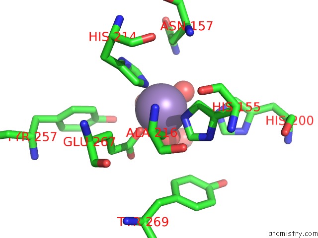
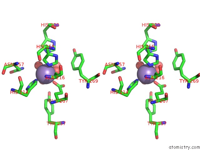
Manganese binding site 1 out of 4 in 3bza
Go back to
Manganese binding site 1 out
of 4 in the Structure of Mn-Substituted Homoprotocatechuate 2,3-Dioxygenase From B.Fuscum at 1.7 Ang Resolution
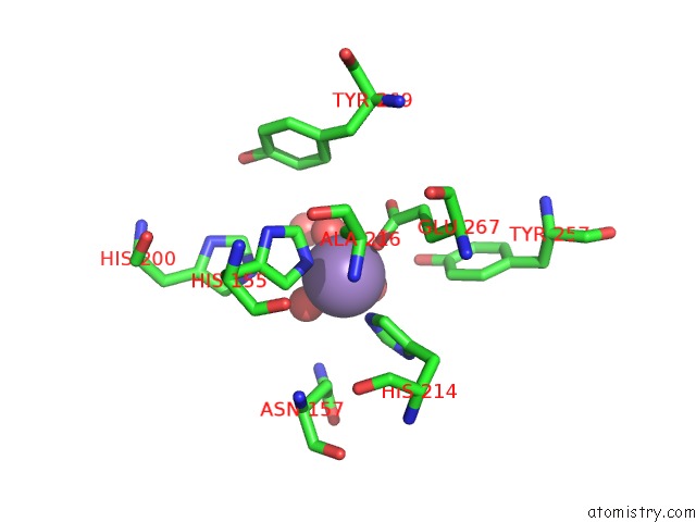
Mono view
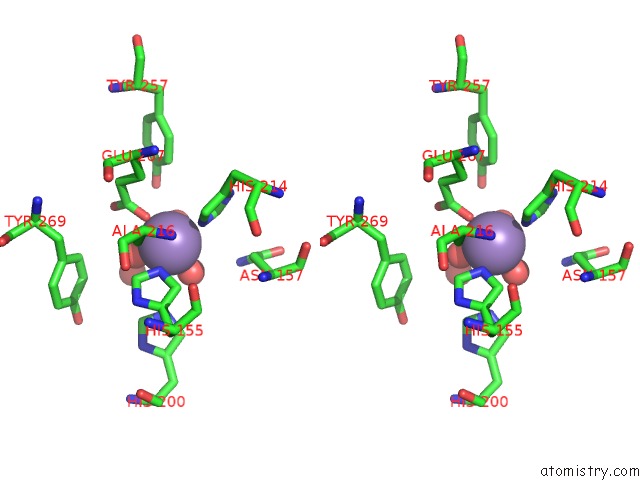
Stereo pair view
Mono view
Stereo pair view
A full contact list of Manganese with other atoms in the Mn binding
site number 1 of Structure of Mn-Substituted Homoprotocatechuate 2,3-Dioxygenase From B.Fuscum at 1.7 Ang Resolution within 5.0Å range:
|
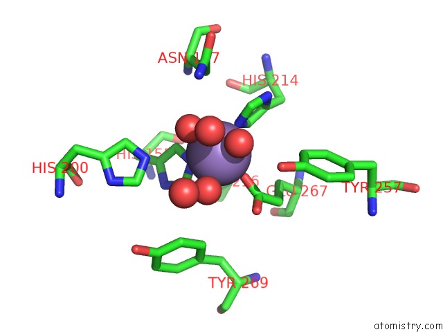
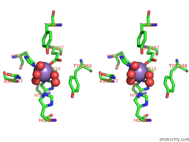
Manganese binding site 2 out of 4 in 3bza
Go back to
Manganese binding site 2 out
of 4 in the Structure of Mn-Substituted Homoprotocatechuate 2,3-Dioxygenase From B.Fuscum at 1.7 Ang Resolution
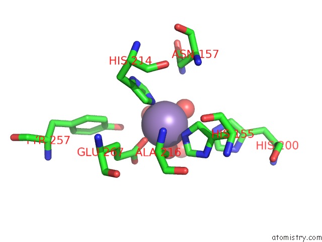
Mono view
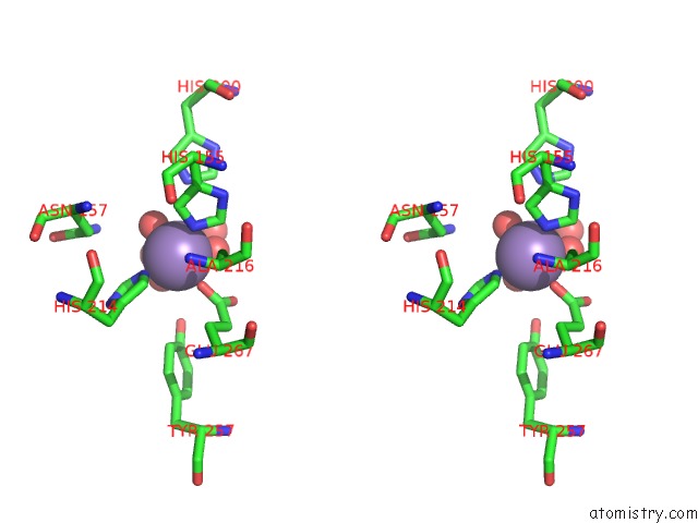
Stereo pair view
Mono view
Stereo pair view
A full contact list of Manganese with other atoms in the Mn binding
site number 2 of Structure of Mn-Substituted Homoprotocatechuate 2,3-Dioxygenase From B.Fuscum at 1.7 Ang Resolution within 5.0Å range:
|
Manganese binding site 3 out of 4 in 3bza
Go back to
Manganese binding site 3 out
of 4 in the Structure of Mn-Substituted Homoprotocatechuate 2,3-Dioxygenase From B.Fuscum at 1.7 Ang Resolution
Mono view
Stereo pair view
Mono view
Stereo pair view
A full contact list of Manganese with other atoms in the Mn binding
site number 3 of Structure of Mn-Substituted Homoprotocatechuate 2,3-Dioxygenase From B.Fuscum at 1.7 Ang Resolution within 5.0Å range:
|
Manganese binding site 4 out of 4 in 3bza
Go back to
Manganese binding site 4 out
of 4 in the Structure of Mn-Substituted Homoprotocatechuate 2,3-Dioxygenase From B.Fuscum at 1.7 Ang Resolution
Mono view
Stereo pair view
Mono view
Stereo pair view
A full contact list of Manganese with other atoms in the Mn binding
site number 4 of Structure of Mn-Substituted Homoprotocatechuate 2,3-Dioxygenase From B.Fuscum at 1.7 Ang Resolution within 5.0Å range:
|
Reference:
J.P.Emerson,
E.G.Kovaleva,
E.R.Farquhar,
J.D.Lipscomb,
L.Que.
Swapping Metals in Fe- and Mn-Dependent Dioxygenases: Evidence For Oxygen Activation Without A Change in Metal Redox State. Proc.Natl.Acad.Sci.Usa V. 105 7347 2008.
ISSN: ISSN 0027-8424
PubMed: 18492808
DOI: 10.1073/PNAS.0711179105
Page generated: Sat Oct 5 15:59:20 2024
ISSN: ISSN 0027-8424
PubMed: 18492808
DOI: 10.1073/PNAS.0711179105
Last articles
Zn in 9MJ5Zn in 9HNW
Zn in 9G0L
Zn in 9FNE
Zn in 9DZN
Zn in 9E0I
Zn in 9D32
Zn in 9DAK
Zn in 8ZXC
Zn in 8ZUF