Manganese »
PDB 3auz-3c5m »
3bie »
Manganese in PDB 3bie: X-Ray Structure of E Coli Alkb Bound to Dsdna Containing 1MEA/T with Mn and 2KG
Protein crystallography data
The structure of X-Ray Structure of E Coli Alkb Bound to Dsdna Containing 1MEA/T with Mn and 2KG, PDB code: 3bie
was solved by
C.Yi,
C.-G.Yang,
with X-Ray Crystallography technique. A brief refinement statistics is given in the table below:
| Resolution Low / High (Å) | 20.00 / 1.68 |
| Space group | P 1 21 1 |
| Cell size a, b, c (Å), α, β, γ (°) | 41.413, 75.822, 52.228, 90.00, 108.53, 90.00 |
| R / Rfree (%) | 18.7 / 21.1 |
Manganese Binding Sites:
The binding sites of Manganese atom in the X-Ray Structure of E Coli Alkb Bound to Dsdna Containing 1MEA/T with Mn and 2KG
(pdb code 3bie). This binding sites where shown within
5.0 Angstroms radius around Manganese atom.
In total only one binding site of Manganese was determined in the X-Ray Structure of E Coli Alkb Bound to Dsdna Containing 1MEA/T with Mn and 2KG, PDB code: 3bie:
In total only one binding site of Manganese was determined in the X-Ray Structure of E Coli Alkb Bound to Dsdna Containing 1MEA/T with Mn and 2KG, PDB code: 3bie:
Manganese binding site 1 out of 1 in 3bie
Go back to
Manganese binding site 1 out
of 1 in the X-Ray Structure of E Coli Alkb Bound to Dsdna Containing 1MEA/T with Mn and 2KG
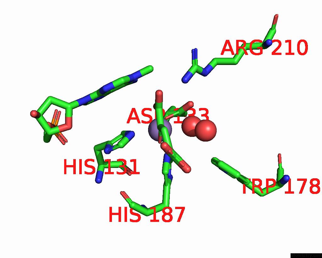
Mono view
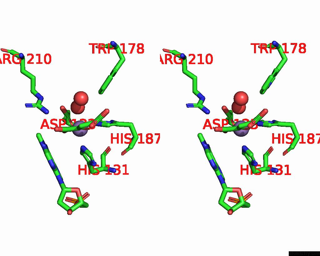
Stereo pair view
Mono view
Stereo pair view
A full contact list of Manganese with other atoms in the Mn binding
site number 1 of X-Ray Structure of E Coli Alkb Bound to Dsdna Containing 1MEA/T with Mn and 2KG within 5.0Å range:
|
Reference:
C.-G.Yang,
C.Yi,
E.M.Duguid,
C.T.Sullivan,
X.Jian,
P.A.Rice,
C.He.
Crystal Structures of Dna/Rna Repair Enzymes Alkb and ABH2 Bound to Dsdna Nature V. 452 961 2008.
ISSN: ISSN 0028-0836
PubMed: 18432238
DOI: 10.1038/NATURE06889
Page generated: Sat Oct 5 15:57:37 2024
ISSN: ISSN 0028-0836
PubMed: 18432238
DOI: 10.1038/NATURE06889
Last articles
Ca in 5SZMCa in 5SZL
Ca in 5SY1
Ca in 5SWI
Ca in 5SVE
Ca in 5SSX
Ca in 5SV0
Ca in 5STD
Ca in 5SSZ
Ca in 5SSY