Manganese »
PDB 3auz-3c5m »
3bfr »
Manganese in PDB 3bfr: The Crystal Structure of SOD2 From Saccharomyces Cerevisiae
Enzymatic activity of The Crystal Structure of SOD2 From Saccharomyces Cerevisiae
All present enzymatic activity of The Crystal Structure of SOD2 From Saccharomyces Cerevisiae:
1.15.1.1;
1.15.1.1;
Protein crystallography data
The structure of The Crystal Structure of SOD2 From Saccharomyces Cerevisiae, PDB code: 3bfr
was solved by
Y.-X.He,
M.-X.Zhao,
C.Zhou,
with X-Ray Crystallography technique. A brief refinement statistics is given in the table below:
| Resolution Low / High (Å) | 15.00 / 2.05 |
| Space group | P 2 3 |
| Cell size a, b, c (Å), α, β, γ (°) | 92.740, 92.740, 92.740, 90.00, 90.00, 90.00 |
| R / Rfree (%) | 19.5 / 23.4 |
Manganese Binding Sites:
The binding sites of Manganese atom in the The Crystal Structure of SOD2 From Saccharomyces Cerevisiae
(pdb code 3bfr). This binding sites where shown within
5.0 Angstroms radius around Manganese atom.
In total only one binding site of Manganese was determined in the The Crystal Structure of SOD2 From Saccharomyces Cerevisiae, PDB code: 3bfr:
In total only one binding site of Manganese was determined in the The Crystal Structure of SOD2 From Saccharomyces Cerevisiae, PDB code: 3bfr:
Manganese binding site 1 out of 1 in 3bfr
Go back to
Manganese binding site 1 out
of 1 in the The Crystal Structure of SOD2 From Saccharomyces Cerevisiae
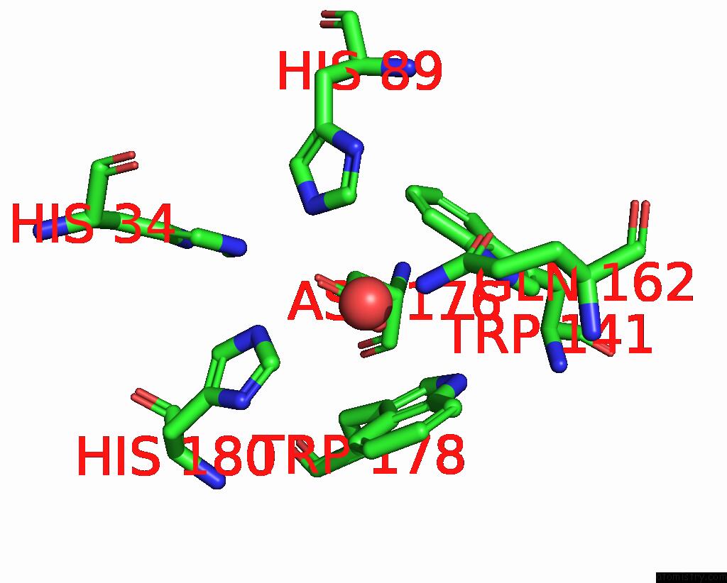
Mono view
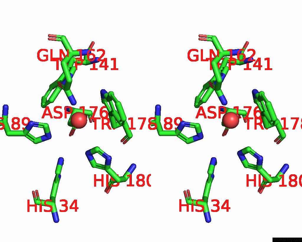
Stereo pair view
Mono view
Stereo pair view
A full contact list of Manganese with other atoms in the Mn binding
site number 1 of The Crystal Structure of SOD2 From Saccharomyces Cerevisiae within 5.0Å range:
|
Reference:
Y.Kang,
Y.-X.He,
M.-X.Zhao,
W.-F.Li.
Structures of Native and Fe-Substituted SOD2 From Saccharomyces Cerevisiae Acta Crystallogr.,Sect.F V. 67 1173 2011.
ISSN: ESSN 1744-3091
PubMed: 22102021
DOI: 10.1107/S1744309111029186
Page generated: Sat Aug 16 11:30:33 2025
ISSN: ESSN 1744-3091
PubMed: 22102021
DOI: 10.1107/S1744309111029186
Last articles
Na in 1R4PNa in 1R8C
Na in 1R8B
Na in 1R8A
Na in 1R89
Na in 1QY1
Na in 1QY2
Na in 1QY0
Na in 1QW7
Na in 1QTK