Manganese »
PDB 2ypq-3au8 »
3a6u »
Manganese in PDB 3a6u: Crystal Structure of Mutt-8-Oxo-Dgmp-Mn(II) Complex
Protein crystallography data
The structure of Crystal Structure of Mutt-8-Oxo-Dgmp-Mn(II) Complex, PDB code: 3a6u
was solved by
T.Nakamura,
Y.Yamagata,
with X-Ray Crystallography technique. A brief refinement statistics is given in the table below:
| Resolution Low / High (Å) | 18.20 / 2.56 |
| Space group | P 21 21 21 |
| Cell size a, b, c (Å), α, β, γ (°) | 38.234, 56.003, 59.339, 90.00, 90.00, 90.00 |
| R / Rfree (%) | 19.3 / 24.2 |
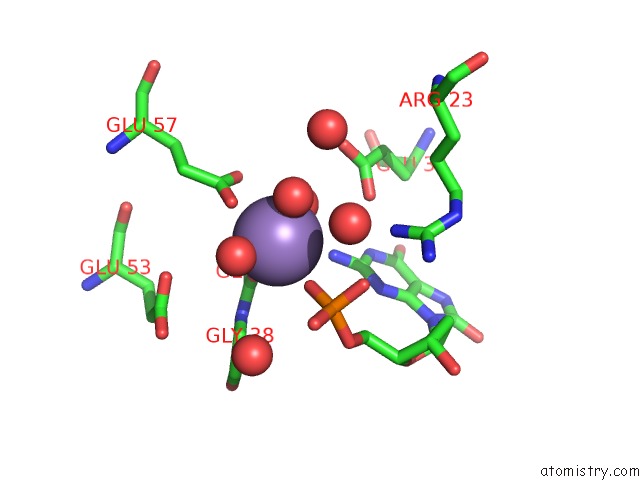
Manganese Binding Sites:
The binding sites of Manganese atom in the Crystal Structure of Mutt-8-Oxo-Dgmp-Mn(II) Complex
(pdb code 3a6u). This binding sites where shown within
5.0 Angstroms radius around Manganese atom.
In total only one binding site of Manganese was determined in the Crystal Structure of Mutt-8-Oxo-Dgmp-Mn(II) Complex, PDB code: 3a6u:
In total only one binding site of Manganese was determined in the Crystal Structure of Mutt-8-Oxo-Dgmp-Mn(II) Complex, PDB code: 3a6u:
Manganese binding site 1 out of 1 in 3a6u
Go back to
Manganese binding site 1 out
of 1 in the Crystal Structure of Mutt-8-Oxo-Dgmp-Mn(II) Complex
Mono view
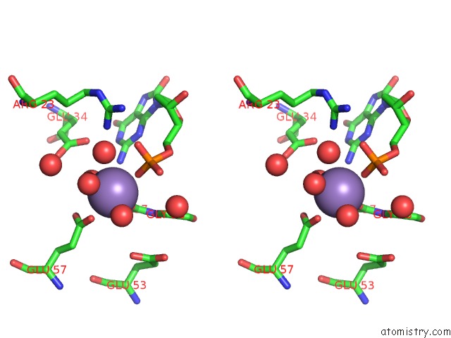
Stereo pair view
Mono view
Stereo pair view
A full contact list of Manganese with other atoms in the Mn binding
site number 1 of Crystal Structure of Mutt-8-Oxo-Dgmp-Mn(II) Complex within 5.0Å range:
|
Reference:
T.Nakamura,
S.Meshitsuka,
S.Kitagawa,
N.Abe,
J.Yamada,
T.Ishino,
H.Nakano,
T.Tsuzuki,
T.Doi,
Y.Kobayashi,
S.Fujii,
M.Sekiguchi,
Y.Yamagata.
Structural and Dynamic Features of the Mutt Protein in the Recognition of Nucleotides with the Mutagenic 8-Oxoguanine Base J.Biol.Chem. V. 285 444 2010.
ISSN: ISSN 0021-9258
PubMed: 19864691
DOI: 10.1074/JBC.M109.066373
Page generated: Sat Oct 5 15:43:38 2024
ISSN: ISSN 0021-9258
PubMed: 19864691
DOI: 10.1074/JBC.M109.066373
Last articles
Zn in 9J0NZn in 9J0O
Zn in 9J0P
Zn in 9FJX
Zn in 9EKB
Zn in 9C0F
Zn in 9CAH
Zn in 9CH0
Zn in 9CH3
Zn in 9CH1