Manganese »
PDB 2ypq-3au8 »
2zbj »
Manganese in PDB 2zbj: Crystal Structure of Dioclea Rostrata Lectin
Protein crystallography data
The structure of Crystal Structure of Dioclea Rostrata Lectin, PDB code: 2zbj
was solved by
T.M.De Oliveira,
P.Delatorre,
B.A.M.Da Rocha,
E.P.De Sousa,
K.S.Nascimento,
G.A.Bezerra,
T.R.Moura,
R.G.Benevides,
E.H.S.Bezerra,
F.B.M.B.Moreno,
V.N.Freire,
W.F.De Azevedo Jr.,
B.S.Cavada,
with X-Ray Crystallography technique. A brief refinement statistics is given in the table below:
| Resolution Low / High (Å) | 33.11 / 2.05 |
| Space group | I 2 2 2 |
| Cell size a, b, c (Å), α, β, γ (°) | 61.511, 88.220, 87.764, 90.00, 90.00, 90.00 |
| R / Rfree (%) | 18.7 / 23.8 |
Other elements in 2zbj:
The structure of Crystal Structure of Dioclea Rostrata Lectin also contains other interesting chemical elements:
| Calcium | (Ca) | 1 atom |
Manganese Binding Sites:
The binding sites of Manganese atom in the Crystal Structure of Dioclea Rostrata Lectin
(pdb code 2zbj). This binding sites where shown within
5.0 Angstroms radius around Manganese atom.
In total only one binding site of Manganese was determined in the Crystal Structure of Dioclea Rostrata Lectin, PDB code: 2zbj:
In total only one binding site of Manganese was determined in the Crystal Structure of Dioclea Rostrata Lectin, PDB code: 2zbj:
Manganese binding site 1 out of 1 in 2zbj
Go back to
Manganese binding site 1 out
of 1 in the Crystal Structure of Dioclea Rostrata Lectin
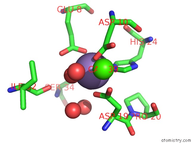
Mono view
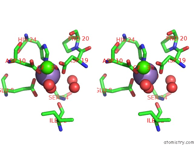
Stereo pair view
Mono view
Stereo pair view
A full contact list of Manganese with other atoms in the Mn binding
site number 1 of Crystal Structure of Dioclea Rostrata Lectin within 5.0Å range:
|
Reference:
T.M.De Oliveira,
P.Delatorre,
B.A.M.Da Rocha,
E.P.De Souza,
K.S.Nascimento,
G.A.Bezerra,
T.R.Moura,
R.G.Benevides,
E.H.S.Bezerra,
F.B.M.B.Moreno,
V.N.Freire,
W.F.De Azevedo Jr.,
B.S.Cavada.
Crystal Structure of Dioclea Rostrata Lectin: Insights Into Understanding the pH-Dependent Dimer-Tetramer Equilibrium and the Structural Basis For Carbohydrate Recognition in Diocleinae Lectins J.Struct.Biol. V. 164 177 2008.
ISSN: ISSN 1047-8477
PubMed: 18682294
DOI: 10.1016/J.JSB.2008.05.012
Page generated: Sat Oct 5 15:34:58 2024
ISSN: ISSN 1047-8477
PubMed: 18682294
DOI: 10.1016/J.JSB.2008.05.012
Last articles
Zn in 9MJ5Zn in 9HNW
Zn in 9G0L
Zn in 9FNE
Zn in 9DZN
Zn in 9E0I
Zn in 9D32
Zn in 9DAK
Zn in 8ZXC
Zn in 8ZUF