Manganese »
PDB 2ypq-3au8 »
2z86 »
Manganese in PDB 2z86: Crystal Structure of Chondroitin Polymerase From Escherichia Coli Strain K4 (K4CP) Complexed with Udp-Glcua and Udp
Enzymatic activity of Crystal Structure of Chondroitin Polymerase From Escherichia Coli Strain K4 (K4CP) Complexed with Udp-Glcua and Udp
All present enzymatic activity of Crystal Structure of Chondroitin Polymerase From Escherichia Coli Strain K4 (K4CP) Complexed with Udp-Glcua and Udp:
2.4.1.175; 2.4.1.226;
2.4.1.175; 2.4.1.226;
Protein crystallography data
The structure of Crystal Structure of Chondroitin Polymerase From Escherichia Coli Strain K4 (K4CP) Complexed with Udp-Glcua and Udp, PDB code: 2z86
was solved by
T.Osawa,
N.Sugiura,
H.Shimada,
R.Hirooka,
A.Tsuji,
M.Kimura,
K.Kimata,
Y.Kakuta,
with X-Ray Crystallography technique. A brief refinement statistics is given in the table below:
| Resolution Low / High (Å) | 20.00 / 2.40 |
| Space group | P 1 21 1 |
| Cell size a, b, c (Å), α, β, γ (°) | 84.092, 219.829, 85.856, 90.00, 103.07, 90.00 |
| R / Rfree (%) | 22.5 / 28.6 |
Manganese Binding Sites:
The binding sites of Manganese atom in the Crystal Structure of Chondroitin Polymerase From Escherichia Coli Strain K4 (K4CP) Complexed with Udp-Glcua and Udp
(pdb code 2z86). This binding sites where shown within
5.0 Angstroms radius around Manganese atom.
In total 8 binding sites of Manganese where determined in the Crystal Structure of Chondroitin Polymerase From Escherichia Coli Strain K4 (K4CP) Complexed with Udp-Glcua and Udp, PDB code: 2z86:
Jump to Manganese binding site number: 1; 2; 3; 4; 5; 6; 7; 8;
In total 8 binding sites of Manganese where determined in the Crystal Structure of Chondroitin Polymerase From Escherichia Coli Strain K4 (K4CP) Complexed with Udp-Glcua and Udp, PDB code: 2z86:
Jump to Manganese binding site number: 1; 2; 3; 4; 5; 6; 7; 8;
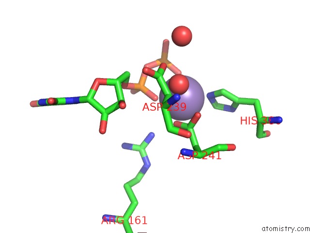
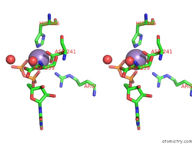
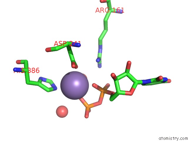
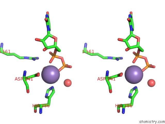
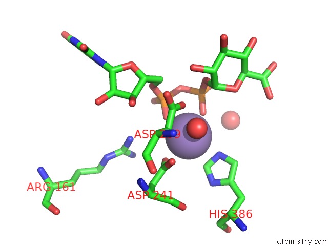
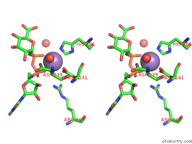
Manganese binding site 1 out of 8 in 2z86
Go back to
Manganese binding site 1 out
of 8 in the Crystal Structure of Chondroitin Polymerase From Escherichia Coli Strain K4 (K4CP) Complexed with Udp-Glcua and Udp
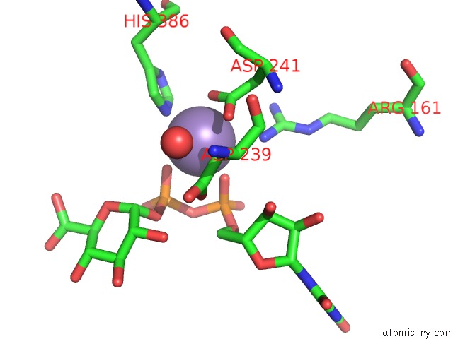
Mono view
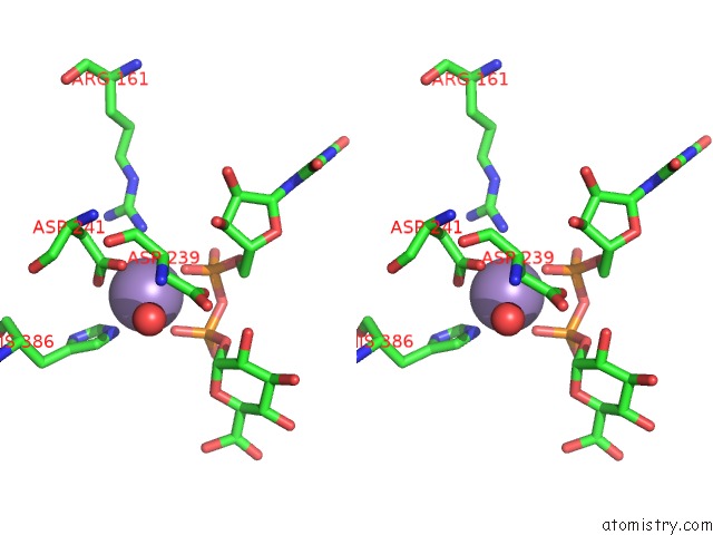
Stereo pair view
Mono view
Stereo pair view
A full contact list of Manganese with other atoms in the Mn binding
site number 1 of Crystal Structure of Chondroitin Polymerase From Escherichia Coli Strain K4 (K4CP) Complexed with Udp-Glcua and Udp within 5.0Å range:
|
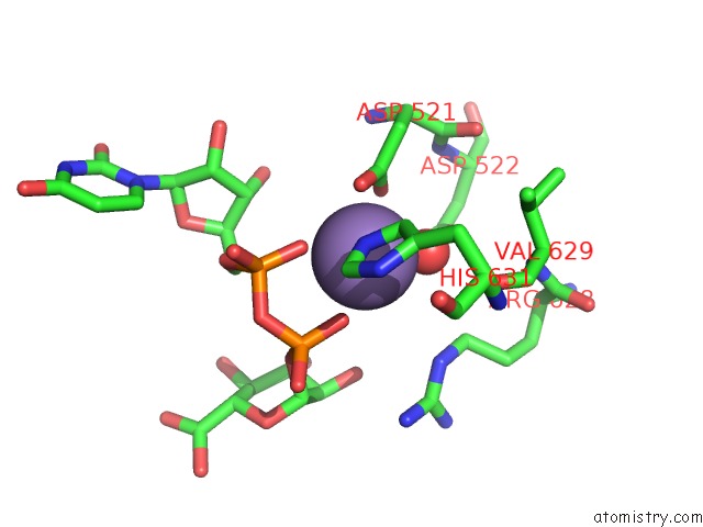
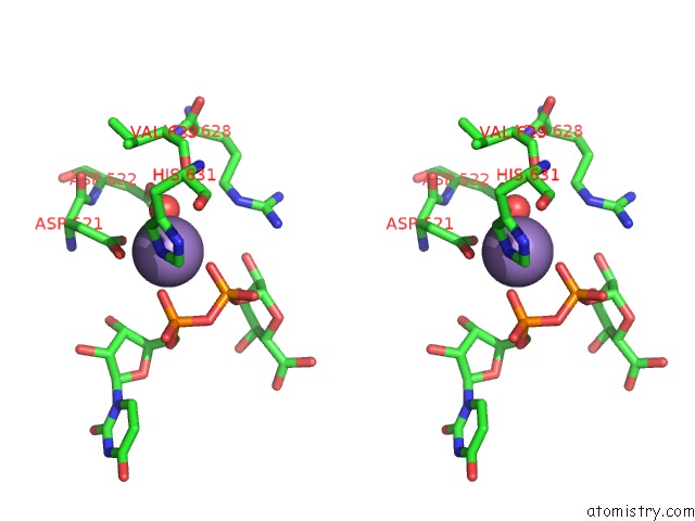
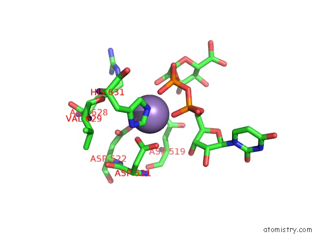
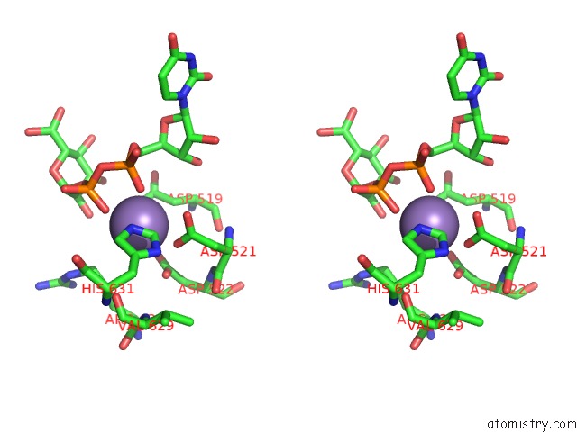
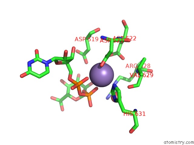
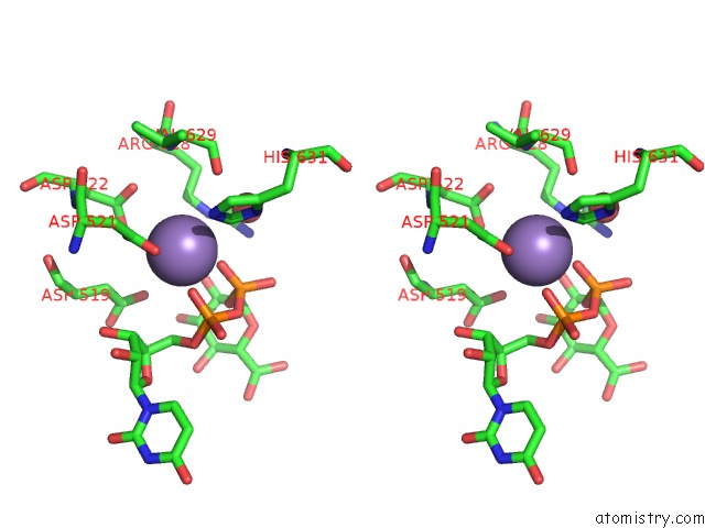
Manganese binding site 2 out of 8 in 2z86
Go back to
Manganese binding site 2 out
of 8 in the Crystal Structure of Chondroitin Polymerase From Escherichia Coli Strain K4 (K4CP) Complexed with Udp-Glcua and Udp
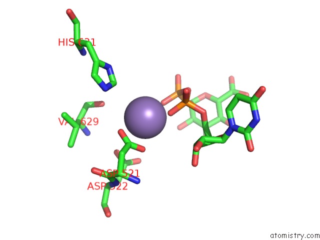
Mono view
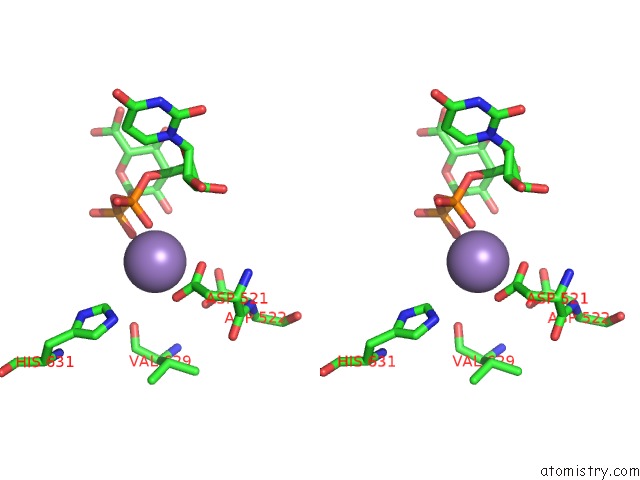
Stereo pair view
Mono view
Stereo pair view
A full contact list of Manganese with other atoms in the Mn binding
site number 2 of Crystal Structure of Chondroitin Polymerase From Escherichia Coli Strain K4 (K4CP) Complexed with Udp-Glcua and Udp within 5.0Å range:
|
Manganese binding site 3 out of 8 in 2z86
Go back to
Manganese binding site 3 out
of 8 in the Crystal Structure of Chondroitin Polymerase From Escherichia Coli Strain K4 (K4CP) Complexed with Udp-Glcua and Udp
Mono view
Stereo pair view
Mono view
Stereo pair view
A full contact list of Manganese with other atoms in the Mn binding
site number 3 of Crystal Structure of Chondroitin Polymerase From Escherichia Coli Strain K4 (K4CP) Complexed with Udp-Glcua and Udp within 5.0Å range:
|
Manganese binding site 4 out of 8 in 2z86
Go back to
Manganese binding site 4 out
of 8 in the Crystal Structure of Chondroitin Polymerase From Escherichia Coli Strain K4 (K4CP) Complexed with Udp-Glcua and Udp
Mono view
Stereo pair view
Mono view
Stereo pair view
A full contact list of Manganese with other atoms in the Mn binding
site number 4 of Crystal Structure of Chondroitin Polymerase From Escherichia Coli Strain K4 (K4CP) Complexed with Udp-Glcua and Udp within 5.0Å range:
|
Manganese binding site 5 out of 8 in 2z86
Go back to
Manganese binding site 5 out
of 8 in the Crystal Structure of Chondroitin Polymerase From Escherichia Coli Strain K4 (K4CP) Complexed with Udp-Glcua and Udp
Mono view
Stereo pair view
Mono view
Stereo pair view
A full contact list of Manganese with other atoms in the Mn binding
site number 5 of Crystal Structure of Chondroitin Polymerase From Escherichia Coli Strain K4 (K4CP) Complexed with Udp-Glcua and Udp within 5.0Å range:
|
Manganese binding site 6 out of 8 in 2z86
Go back to
Manganese binding site 6 out
of 8 in the Crystal Structure of Chondroitin Polymerase From Escherichia Coli Strain K4 (K4CP) Complexed with Udp-Glcua and Udp
Mono view
Stereo pair view
Mono view
Stereo pair view
A full contact list of Manganese with other atoms in the Mn binding
site number 6 of Crystal Structure of Chondroitin Polymerase From Escherichia Coli Strain K4 (K4CP) Complexed with Udp-Glcua and Udp within 5.0Å range:
|
Manganese binding site 7 out of 8 in 2z86
Go back to
Manganese binding site 7 out
of 8 in the Crystal Structure of Chondroitin Polymerase From Escherichia Coli Strain K4 (K4CP) Complexed with Udp-Glcua and Udp
Mono view
Stereo pair view
Mono view
Stereo pair view
A full contact list of Manganese with other atoms in the Mn binding
site number 7 of Crystal Structure of Chondroitin Polymerase From Escherichia Coli Strain K4 (K4CP) Complexed with Udp-Glcua and Udp within 5.0Å range:
|
Manganese binding site 8 out of 8 in 2z86
Go back to
Manganese binding site 8 out
of 8 in the Crystal Structure of Chondroitin Polymerase From Escherichia Coli Strain K4 (K4CP) Complexed with Udp-Glcua and Udp
Mono view
Stereo pair view
Mono view
Stereo pair view
A full contact list of Manganese with other atoms in the Mn binding
site number 8 of Crystal Structure of Chondroitin Polymerase From Escherichia Coli Strain K4 (K4CP) Complexed with Udp-Glcua and Udp within 5.0Å range:
|
Reference:
T.Osawa,
N.Sugiura,
H.Shimada,
R.Hirooka,
A.Tsuji,
T.Shirakawa,
K.Fukuyama,
M.Kimura,
K.Kimata,
Y.Kakuta.
Crystal Structure of Chondroitin Polymerase From Escherichia Coli K4. Biochem. Biophys. Res. V. 378 10 2009COMMUN..
ISSN: ESSN 1090-2104
PubMed: 18771653
DOI: 10.1016/J.BBRC.2008.08.121
Page generated: Sat Oct 5 15:34:59 2024
ISSN: ESSN 1090-2104
PubMed: 18771653
DOI: 10.1016/J.BBRC.2008.08.121
Last articles
Ca in 5VS2Ca in 5VOL
Ca in 5VRX
Ca in 5VRW
Ca in 5VR0
Ca in 5VPL
Ca in 5VPH
Ca in 5VMS
Ca in 5VPG
Ca in 5VOF